When Willem Johan Kolff invented dialysis, the "father" of artificial organs was just getting started.
One of the Netherlands’ most famous pieces of pop culture is “Soldier of Orange.” It’s the title of the country’s most celebrated war memoir, movie and epic stage musical, all of which detail the exploits of the nation’s resistance fighters during World War II.
Willem Johan Kolff was a member of the Dutch resistance, but he doesn’t rate a mention in the “Solider of Orange” canon. Yet his wartime toils in a rural backwater not only changed medicine, but the world.
Kolff had been a physician less than two years before Germany invaded the Netherlands in May 1940. He had been engaged in post-graduate studies at the University of Gronigen but withdrew because he refused to accommodate the demands of the Nazi occupiers. Kolff’s Jewish supervisor made an even starker choice: He committed suicide.
After his departure from the university, Kolff took a job managing a small hospital in Kampen. Located 50 miles from the heavily populated coastal region, the facility was far enough away from the prying eyes of Germans that not only could Kolff care for patients, he could hide fellow resistance fighters and even Jewish refugees in relative safety. Kolff coached many of them to feign convincing terminal illnesses so the Nazis would allow them to remain in the hospital.
Despite the demands of practicing medicine and resistance work, Kolff still found time to conduct research. He had been haunted and inspired when, not long before the Nazi invasion, one of his patients died in agony from kidney disease. Kolff wanted to find a way to save future patients.
He broke his problem down to a simple task: If he could remove 20 grams of urea from a patient’s blood in 24 hours, they would survive. He began experimenting with ways to filter blood and return it to a patient’s body. Since the war had ground all non-military manufacturing to a halt, he was mostly forced to make do with material he could find at the hospital and around Kampen. Kolff eventually built a device from a washing machine parts, juice cans, sausage casings, a valve from an old Ford automobile radiator, and even scrap from a downed German aircraft.
The world’s first dialysis machine was hardly imposing; it resembled a rotating drum for a bingo game or raffle. Yet it carried on the highly sophisticated task of moving a patient’s blood through a semi-permeable membrane (about a 50-foot length of sausage casings) into a saline solution that drew out urea while leaving the blood cells untouched.
In emigrating to the U.S. to practice medicine, Kolff's intent was twofold: Advocate for a wider adoption of dialysis, and work on new projects. He wildly succeeded at both.
Kolff began using the machine to treat patients in 1943, most of whom had lapsed into comas due to their kidney failure. But like most groundbreaking medical devices, it was not an immediate success. By the end of the war, Kolff had dialyzed more than a dozen patients, but all had died. He briefly suspended use of the device after the Allied invasion of Europe, but he continued to refine its operation and the administration of blood thinners to patients.
In September 1945, Kolff dialyzed another comatose patient, 67-year-old Sofia Maria Schafstadt. She regained consciousness after 11 hours, and would live well into the 1950s with Kolff’s assistance. Yet this triumph contained a dark irony: At the time of her treatment, Schafstadt had been imprisoned for collaborating with the Germans.
With a tattered Europe struggling to overcome the destruction of the war, Kolff and his family emigrated to the U.S. in 1950, where he began working for the Cleveland Clinic while undergoing the naturalization process so he could practice medicine in the U.S. His intent was twofold: Advocate for a wider adoption of dialysis, and work on new projects. He wildly succeeded at both.
By the mid-1950s, dialysis machines had become reliable and life-saving medical devices, and Kolff had become a U.S. citizen. About that time he invented a membrane oxygenator that could be used in heart bypass surgeries. This was a critical component of the heart-lung machine, which would make heart transplants possible and bypass surgeries routine. He also invented among the very first practical artificial hearts, which in 1957 kept a dog alive for 90 minutes.
Kolff moved to the University of Utah in 1967 to become director of its Institute for Biomedical Engineering. It was a promising time for such a move, as the first successful transplant of a donor heart to a human occurred that year. But he was interested in going a step further and creating an artificial heart for human use.
It took more than a decade of tinkering and research, but in 1982, a team of physicians and engineers led by Kolff succeeded in implanting the first artificial heart in dentist Barney Clark, whose failing health disqualified him from a heart transplant. Although Clark died in March 1983 after 112 days tethered to the device, that it kept him alive generated international headlines. While graduate student Robert Jarvik received the named credit for the heart, he was directly supervised by Kolff, whose various endeavors into artificial organ research at the University of Utah were segmented into numerous teams.
Forty years later, several artificial hearts have been approved for use by the Food and Drug Administration, although all are a “bridge” that allow patients to wait for a transplant.
Kolff continued researching and tinkering with biomedical devices – including artificial eyes and ears – until he retired in 1997 at the age of 86. When he died in 2009, the medical community acknowledged that he was not only a pioneer in biotechnology, but the “father” of artificial organs.
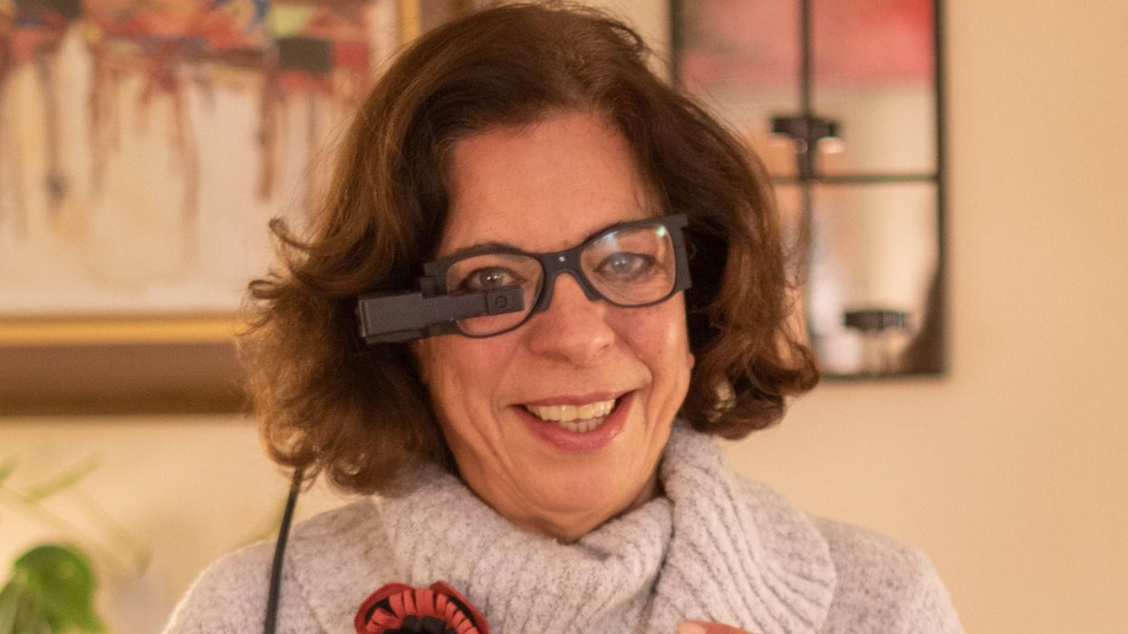
An implant, combined with the glasses and tiny video camera modeled in this photo, could improve the eyesight of millions of people with degenerative eye diseases in the coming years.
For millions of people with macular degeneration, treatment options are slim. The disease causes loss of central vision, which allows us to see straight ahead, and is highly dependent on age, with people over 75 at approximately 30% risk of developing the disorder. The BrightFocus Foundation estimates 11 million people in the U.S. currently have one of three forms of the disease.
Recently, ophthalmologists including Daniel Palanker at Stanford University published research showing advances in the PRIMA retinal implant, which could help people with advanced, age-related macular degeneration regain some of their sight. In a feasibility study, five patients had a pixelated chip implanted behind the retina, and three were able to see using their remaining peripheral vision and—thanks to the implant—their partially restored central vision at the same time.
Should people with macular degeneration be excited about these results?
“Every week, if not every day, patients come to me with this question because it's devastating when they lose their central vision,” says retinal surgeon Lynn Huang. About 40% of her patients have macular degeneration. Huang tells them that these implants, along with new medications and stem cell therapies, could be useful in the coming years.
“The goal here is to replace the missing photoreceptors with photovoltaic pixels, basically like little solar panels,” Palanker says.
That implant, a pixelated chip, works together with a tiny video camera on a specially designed pair of eyeglasses, which can be adjusted for each patient’s prescription. The video camera relays processed images to the chip, which electrically stimulates inner retinal neurons. These neurons, in turn, relay information to the brain’s visual cortex through the optic nerve. The chip restores patients’ central sight, but not completely. The artificial vision is basically monochromatic (whitish-yellowish) and fairly blurry; patients were still legally blind even after the implant, except when using a zoom function on the camera, but those with proper chip placement could make out large letters.
“The goal here is to replace the missing photoreceptors with photovoltaic pixels, basically like little solar panels,” Palanker says. These pixels, located on the implanted chip, convert light into pulsed electrical currents that stimulate retinal neurons. In time, Palanker hopes to improve the chips, resulting in bigger boosts to visual acuity.
The pixelated chips are surgically implanted during a process Palanker admits is still “a surgical learning curve.” In the study, three chips were implanted correctly, one was placed incorrectly, and another patient’s chip moved after the procedure; he did not follow post-surgical recommendations. One patient passed away during the study for unrelated reasons.
University of Maryland retinal specialist Kenneth Taubenslag, who was not involved in the study, said that subretinal surgeries have become less common in recent years, but expects implants to spur improvements in these techniques. “I think as people get more experience, [they’ll] probably get more reliable placement of the implant,” he said, pointing out that even the patient with the misplaced chip was able to gain some light perception, if not the same visual acuity as other patients.
Retinal implants have come under scrutiny lately. IEEE Spectrum reported that Second Sight, manufacturer of the Argus II implant used for people with retinitis pigmentosa, a genetic disease that causes vision loss, would no longer support the product. After selling hundreds of the implants at $150,000 apiece, company leaders announced they’d “decided to pursue an orderly wind down” of Second Sight in March 2020 in the wake of financial issues. Last month, the company announced a merger, shifting its focus to a new retinal implant, raising questions for patients who have Argus II implants.
Retinal surgeon Eugene de Juan of the University of California, San Francisco, was involved with early studies of the Argus implants, though his participation ended over a decade ago, before the device was marketed by Second Sight. He says he would consider recommending future implants to patients with macular degeneration, given the promise of the technology and the lack of other alternatives.
“I tell my patients that this is an area of active research and development, and it's getting better and better, so let's not give up hope,” de Juan says. He believes cautious optimism for Palanker’s implant is appropriate: “It's not the first, it's not the only, but it's a good approach with a good team.”
New Cell Therapies Give Hope to Diabetes Patients
Researchers are developing new cell therapies that could cure Type 1 diabetes and make constant management of the disease a way of the past.
For nearly four decades, George Huntley has thought constantly about his diabetes. Diagnosed in 1983 with Type 1 (insulin-dependent) diabetes, Huntley began managing his condition with daily finger sticks to check his blood glucose levels and doses of insulin that he injected into his abdomen. Even now, with an insulin pump and a device that continuously monitors his glucose, he must consider how every meal will affect his blood sugar, checking his monitor multiple times each hour.
Like many of those who depend on insulin injections, Huntley is simultaneously grateful for the technology that makes his condition easier to manage and tired of thinking about diabetes. If he could wave a magic wand, he says, he would make his diabetes disappear. So when he read about biotechs like ViaCyte and Vertex Pharmaceuticals developing new cell therapies that have the potential to cure Type 1 diabetes, Huntley was excited.
You also won’t see him signing up any time soon. The therapies under development by both companies would require a lifelong regimen of drugs for suppressing the immune system to prevent the body from rejecting the foreign cells. It’s a problem also seen in the transplant of insulin-producing cells of the pancreas – called islet cells – from deceased donors. To Howard Foyt, chief medical officer at ViaCyte, a San Diego-based biotech specializing in the development of cell therapies for diabetes, the tradeoff is worth it.
“A lot of the symptoms of diabetes are not something that you wear on your arm, so to speak. You’re not necessarily conscious of them until you’re successfully treated, and you feel better,” Foyt says.
For many with diabetes, managing these symptoms is a constant game of Whack-a-Mole. “Any form of treatment that gets someone closer to feeling good is a victory,” he says.
“Am I going to be trading diabetes for cancer? That’s not a chance I
want to take."
But not everyone is convinced. What’s more, it’s likely that the availability of these cell therapies will be limited to those with life-threatening diabetes symptoms, such as hypoglycemia unawareness. To Huntley, these therapies remain a bit of a Faustian bargain.
“Am I going to be trading diabetes for cancer? That’s not a chance I want to take,” he says.
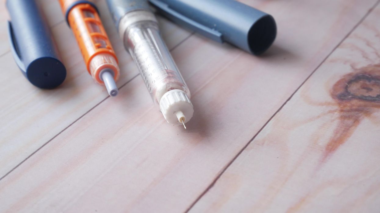
The discovery of insulin in 1921 transformed Type 1 diabetes from a death sentence into a potentially manageable condition. Even as better versions of insulin hit the market—ones that weren’t derived from pigs and wouldn’t provoke an allergic response, longer-acting insulin, insulin pens—they didn’t change the reality that those with Type 1 diabetes remained dependent on insulin. Even the most advanced continuous glucose monitors (which tests blood sugar levels every few minutes, 24/7) and insulin pumps don’t perform as well as a healthy pancreas.
Whether by injection or pump, someone with diabetes needs to administer the insulin their body no longer makes. With advances in organ transplantation, the concept of transplanting insulin-producing pancreatic beta cells seemed obvious. After more than a decade of painstaking work, James Shapiro, who directs the Islet Transplant Program at the University of Albania, honed a process called the Edmonton Protocol for pancreas transplants. For a few patients who couldn’t control their blood sugars any other way, the Edmonton Protocol became a life saver. Some of these patients were even able to stop insulin completely, Shapiro says. But the high cost of organ transplant and a chronic shortage of donor organs, pancreas or otherwise, meant that only a small handful of patients could benefit.
Stem cells, however, can be grown in vats, meaning that supply would never be an issue. “We would be going from a very successful treatment of today to a potential cure tomorrow,” Shapiro says.
In 2014, spurred by his own children’s diagnoses with Type 1 Diabetes, stem cell biologist Doug Melton of Harvard University figured out a way to differentiate embryonic stem cells into functional pancreatic beta cells. It was a long process, explains immunoengineer Alice Tomei at the University of Miami, because “the islet is not one cell, it's like a mini-organ that has its own needs.”
Add on the risk of rejection and autoimmunity, and Tomei says that scientists soon realized that chronic and systemic immunosuppression was the only way forward. Over the next several years, Melton improved his approach to yield more cells with fewer impurities. Melton partnered with Boston-based Vertex Pharmaceuticals to create a cell therapy called VX-880.
The first patient received his dose earlier in 2021. In October, Vertex released 90-day results from the Phase 1/2 trial, which revealed the patient was able to reduce his insulin usage from an average of 34 units per day to just 2.9 units per day. The tradeoff is a lifelong need for immunosuppressive drugs to prevent the body from attacking both foreign cells and pancreatic beta cells. It’s what recipients of ViaCyte’s first-gen PEC-Direct will also need. For Foyt, it’s an easy choice.
“At this point in time, immunosuppression is the necessary evil,” he says. “For parents, would you like to worry about going into your child’s bedroom every morning and not knowing if they’re going to be alive or dead? It’s uncommon, but it does occur.”
Not everyone, however, finds the trade-off easy to swallow. Especially with COVID-19 cases reaching record highs, the prospect of reducing his immune function at a time when he needs it most doesn’t sit well with Huntley. The risks of immunosuppression also mean that diabetes cell therapies are limited to those patients with life-threatening complications.
It’s why ViaCyte has created two new iterations of cellular therapies that would eliminate this need. The ViaCyte-Encap contains the cells in a permeable container that allows oxygen, insulin, and nutrients to flow freely but prevents immune system access. Their latest model, PEC-QT, just began safety trials with Shapiro’s lab at the University of Alberta and uses gene editing to eliminate any cellular markers that would trigger an immune response.
Sanjoy Dutta, vice president of research at JDRF International, a nonprofit that funds the study of diabetes, is thrilled with the progress that’s been made around cell therapies, but he cautions it’s still early days. “We have proven that these cells can be made. What we haven’t seen is are they going to work for six months, two years, five years? It’s a challenge we still need to overcome,” he says.
Iowa social worker Jodi Lynn’s concerns echo Dutta’s. Lynn was diagnosed with diabetes in 1998 at age 14 after a bout of severe influenza, spends each day inventorying supplies, planning her food intake, and maintaining her insulin pump and glucose monitor. These newer technologies dramatically improved her blood sugar control but, like everyone with diabetes, Lynn remains at high risk for complications, such as diabetic ketoacidosis, heart disease, vision loss, and kidney failure. Lynn, already considered immunocompromised due to medications she takes for another autoimmune condition, is less concerned with immune suppression than the untested nature of these therapies.
“I want to know that they will work long-term,” she says.


