Neuromarketers Are Studying Brain Scans to Influence Our Product Choices

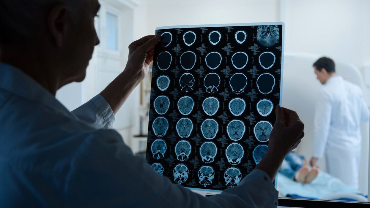
A doctor looking at MRI scan results.
When was the last time you made a pro-con list? Carefully considered all factors and weighed them against each other before you made a choice?
Chances are that most of your decisions do not follow this rigorous process. They are made quickly, subconsciously, and often do not adhere to any strict logic. Rather, your decisions are influenced by your mood, your relatives and friends, and a range of other factors that scientists are still unraveling.
When the shoppers were asked why they chose that bottle of wine, almost none of them noticed the music or believed it influenced their decision.
Influencing your choices is also the holy grail of marketing. Companies spend vast amounts of time and money creating product designs and ads. These ads are often tested in focus groups or individual interviews to ensure that they will do well in the market.
Traditional methods of market research rely on self-reports. The participants are asked which ad they find more appealing and why. But there are a few problems with this approach.
For one, the participants might not fully understand their true preferences. They might think that the green design looks more appealing when they compare choices, but then pick up the orange one when they mindlessly wander through the supermarket. It's well known that we humans often do not act rationally, so why would we accurately predict our own behavior?
Another issue is that we like to think of ourselves as logical. Even though our choices are at least partially made subconsciously, we have a tendency to rationalize them after the fact. For example, when supermarkets play French music, the shoppers are 3-4 times more likely to buy French wine. Play German music and German wine sales go up. But when the shoppers are asked why they chose that bottle of wine, almost none of them notice the music or believe it influenced their decision. Instead, they say that they preferred the label or price.
Finally, participants might truly know their preference but choose not to disclose it. Imagine sitting in a focus group watching a TV spot that makes fun of somebody's misfortune. You might be too embarrassed to admit that this is the funnier and more appealing spot, because you're afraid of being judged.
Results from traditional market research are therefore unavoidably subjective and biased.
In the hope of overcoming these limitations, newer ways of market research have been developed, among them neuromarketing, which applies neuroscience to marketing.
Today, neuromarketers focus their efforts on three main stages: to aid product ideation, evaluate the finished product or prototypes, and develop the best marketing strategy. In all cases, they want to find the option with the most "favorable" brain response – but exactly how this brain response is defined varies vastly between studies.
Perhaps the most promising of all non-traditional techniques is functional magnetic resonance imaging (fMRI). This neuroimaging technique measures brain activity indirectly by tracking changes in blood flow. In short, active brain areas receive more oxygen-rich blood. The fMRI scanner picks up the difference between oxygen-rich and oxygen-poor blood and can therefore measure which brain areas are more active than others. But is there truly an untapped potential in the human brain that can be unlocked using neuroimaging?
A number of studies claim that functional neuroimaging has been successfully applied to marketing scenarios. For example, when researchers tried to predict the success of 6 different ads for chocolate bars, the brain response of 18 women was reportedly more predictive than their self-reported preference. The ad that was rated best in interviews was actually the least successful in a real supermarket. In contrast, the neuroimaging algorithm correctly predicted the top two selling ads.
One of the biggest fears is that the potential insights from neuromarketing studies could be used in new, disturbing ways for consumer manipulation.
This study has a number of limitations, which are representative of the majority of neuromarketing research. The field is full of experiments that are conducted with small samples or using suboptimal protocols, with a lack of appropriate control conditions. While a small number of academic researchers are using rigorous protocols, most studies are conducted by neuromarketing companies or funded by the corporations whose products were tested. Such set-ups raise the risk of biased reporting, calling into question the reliability of the findings. Publication bias – the tendency to publish only positive results which leads to a skewing of reported results in the literature – is especially common for industry-funded studies.
One of the biggest fears is that the potential insights from neuromarketing studies could be used in new, disturbing ways for consumer manipulation. If a new product or ad campaign is designed to target our subconscious decision-making better than ever before, are we less able to resist the purchase? We might believe that we all have a healthy amount of self-control, but when we're in the supermarket after a stressful day or we're struggling to manage the self-control of someone else, like a small child, is it ethical for corporations to tap our unconscious decision-making?
As with any technology, the deciding factor is how it will be used. While there are many dangerous applications that might make unhealthy products one day impossible to resist, there are also some more optimistic scenarios. For example, brain scans have been used to predict the success of an antismoking campaign. If such public health interventions that are notoriously ineffective could encourage more people to make healthier lifestyle choices, don't we all benefit? Or is this still a step too far toward manipulation and propaganda?
The conduct of the studies themselves is another problematic area. Academic researchers must go through a rigorous process before they can start a study, which involves review by an ethics board. In contrast, there are barely any regulations for corporate studies. This is not only relevant for the experience of the participants, but also for how the data are being used. Take an extreme case – the brain scan reveals that the participant has a tumor. Universities have protocols in place for how to deal with these situations – often, the scans would be reviewed by a neuro-radiologist and the participant would be informed. Commercial organizations are under no such obligation.
Neuromarketing carries great potential to nudge positive behavioral change, though it also carries the risk of abuse.
Neuromarketing is now a highly competitive field with many different vendors. The Advertising Research Foundation compared 8 vendors that used neuroscientific methods or biometrics for the research of ad campaigns and found that there were differences in methodology and approach; most were proprietary and vendors were not willing to disclose what they measured and how. This lack of transparency is slowing down progress, as researchers cannot contrast and compare different approaches to optimize them.
Despite these methodological challenges, neuromarketing carries great potential to nudge positive behavioral change, though it also carries the risk of abuse. Where one ends and the other starts will need to be clearly defined. It's time to start a public debate now to inform future laws and regulations for the neuromarketing industry, as these technologies will eventually affect us all.
A new type of cancer therapy is shrinking deadly brain tumors with just one treatment
MRI scans after a new kind of immunotherapy for brain cancer show remarkable progress in one patient just days after the first treatment.
Few cancers are deadlier than glioblastomas—aggressive and lethal tumors that originate in the brain or spinal cord. Five years after diagnosis, less than five percent of glioblastoma patients are still alive—and more often, glioblastoma patients live just 14 months on average after receiving a diagnosis.
But an ongoing clinical trial at Mass General Cancer Center is giving new hope to glioblastoma patients and their families. The trial, called INCIPIENT, is meant to evaluate the effects of a special type of immune cell, called CAR-T cells, on patients with recurrent glioblastoma.
How CAR-T cell therapy works
CAR-T cell therapy is a type of cancer treatment called immunotherapy, where doctors modify a patient’s own immune system specifically to find and destroy cancer cells. In CAR-T cell therapy, doctors extract the patient’s T-cells, which are immune system cells that help fight off disease—particularly cancer. These T-cells are harvested from the patient and then genetically modified in a lab to produce proteins on their surface called chimeric antigen receptors (thus becoming CAR-T cells), which makes them able to bind to a specific protein on the patient’s cancer cells. Once modified, these CAR-T cells are grown in the lab for several weeks so that they can multiply into an army of millions. When enough cells have been grown, these super-charged T-cells are infused back into the patient where they can then seek out cancer cells, bind to them, and destroy them. CAR-T cell therapies have been approved by the US Food and Drug Administration (FDA) to treat certain types of lymphomas and leukemias, as well as multiple myeloma, but haven’t been approved to treat glioblastomas—yet.
CAR-T cell therapies don’t always work against solid tumors, such as glioblastomas. Because solid tumors contain different kinds of cancer cells, some cells can evade the immune system’s detection even after CAR-T cell therapy, according to a press release from Massachusetts General Hospital. For the INCIPIENT trial, researchers modified the CAR-T cells even further in hopes of making them more effective against solid tumors. These second-generation CAR-T cells (called CARv3-TEAM-E T cells) contain special antibodies that attack EFGR, a protein expressed in the majority of glioblastoma tumors. Unlike other CAR-T cell therapies, these particular CAR-T cells were designed to be directly injected into the patient’s brain.
The INCIPIENT trial results
The INCIPIENT trial involved three patients who were enrolled in the study between March and July 2023. All three patients—a 72-year-old man, a 74-year-old man, and a 57-year-old woman—were treated with chemo and radiation and enrolled in the trial with CAR-T cells after their glioblastoma tumors came back.
The results, which were published earlier this year in the New England Journal of Medicine (NEJM), were called “rapid” and “dramatic” by doctors involved in the trial. After just a single infusion of the CAR-T cells, each patient experienced a significant reduction in their tumor sizes. Just two days after receiving the infusion, the glioblastoma tumor of the 72-year-old man decreased by nearly twenty percent. Just two months later the tumor had shrunk by an astonishing 60 percent, and the change was maintained for more than six months. The most dramatic result was in the 57-year-old female patient, whose tumor shrank nearly completely after just one infusion of the CAR-T cells.
The results of the INCIPIENT trial were unexpected and astonishing—but unfortunately, they were also temporary. For all three patients, the tumors eventually began to grow back regardless of the CAR-T cell infusions. According to the press release from MGH, the medical team is now considering treating each patient with multiple infusions or prefacing each treatment with chemotherapy to prolong the response.
While there is still “more to do,” says co-author of the study neuro-oncologist Dr. Elizabeth Gerstner, the results are still promising. If nothing else, these second-generation CAR-T cell infusions may someday be able to give patients more time than traditional treatments would allow.
“These results are exciting but they are also just the beginning,” says Dr. Marcela Maus, a doctor and professor of medicine at Mass General who was involved in the clinical trial. “They tell us that we are on the right track in pursuing a therapy that has the potential to change the outlook for this intractable disease.”
A recent study in The Lancet Oncology showed that AI found 20 percent more cancers on mammogram screens than radiologists alone.
Since the early 2000s, AI systems have eliminated more than 1.7 million jobs, and that number will only increase as AI improves. Some research estimates that by 2025, AI will eliminate more than 85 million jobs.
But for all the talk about job security, AI is also proving to be a powerful tool in healthcare—specifically, cancer detection. One recently published study has shown that, remarkably, artificial intelligence was able to detect 20 percent more cancers in imaging scans than radiologists alone.
Published in The Lancet Oncology, the study analyzed the scans of 80,000 Swedish women with a moderate hereditary risk of breast cancer who had undergone a mammogram between April 2021 and July 2022. Half of these scans were read by AI and then a radiologist to double-check the findings. The second group of scans was read by two researchers without the help of AI. (Currently, the standard of care across Europe is to have two radiologists analyze a scan before diagnosing a patient with breast cancer.)
The study showed that the AI group detected cancer in 6 out of every 1,000 scans, while the radiologists detected cancer in 5 per 1,000 scans. In other words, AI found 20 percent more cancers than the highly-trained radiologists.

But even though the AI was better able to pinpoint cancer on an image, it doesn’t mean radiologists will soon be out of a job. Dr. Laura Heacock, a breast radiologist at NYU, said in an interview with CNN that radiologists do much more than simply screening mammograms, and that even well-trained technology can make errors. “These tools work best when paired with highly-trained radiologists who make the final call on your mammogram. Think of it as a tool like a stethoscope for a cardiologist.”
AI is still an emerging technology, but more and more doctors are using them to detect different cancers. For example, researchers at MIT have developed a program called MIRAI, which looks at patterns in patient mammograms across a series of scans and uses an algorithm to model a patient's risk of developing breast cancer over time. The program was "trained" with more than 200,000 breast imaging scans from Massachusetts General Hospital and has been tested on over 100,000 women in different hospitals across the world. According to MIT, MIRAI "has been shown to be more accurate in predicting the risk for developing breast cancer in the short term (over a 3-year period) compared to traditional tools." It has also been able to detect breast cancer up to five years before a patient receives a diagnosis.
The challenges for cancer-detecting AI tools now is not just accuracy. AI tools are also being challenged to perform consistently well across different ages, races, and breast density profiles, particularly given the increased risks that different women face. For example, Black women are 42 percent more likely than white women to die from breast cancer, despite having nearly the same rates of breast cancer as white women. Recently, an FDA-approved AI device for screening breast cancer has come under fire for wrongly detecting cancer in Black patients significantly more often than white patients.
As AI technology improves, radiologists will be able to accurately scan a more diverse set of patients at a larger volume than ever before, potentially saving more lives than ever.

