Parkinson’s Disease Destroyed My Life. Then I Tried Deep Brain Stimulation.

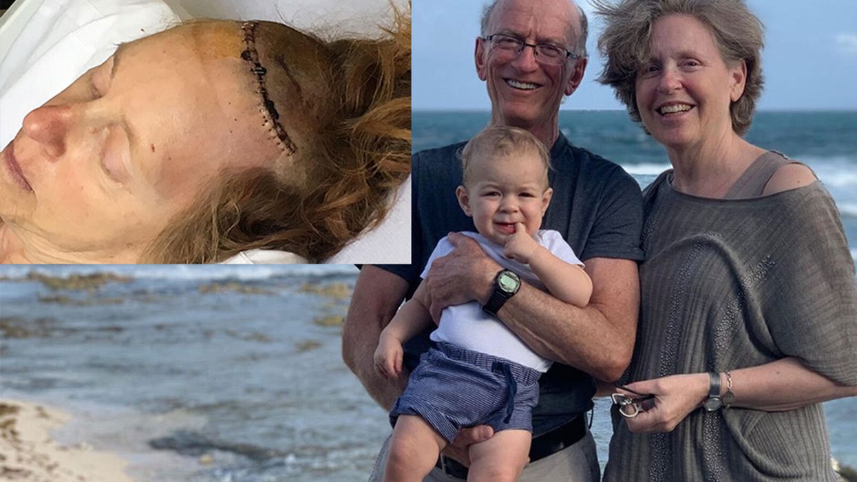
Anne, Stan, and grandson Louie during vacation in Mexico, 2019. INSET: Anne post-op in 2017.
[Editor's Note: On June 6, 2017, Anne Shabason, an artist, hospice educator, and mother of two from Bolton, Ontario, a small town about 30 miles outside of Toronto, underwent Deep Brain Stimulation (DBS) to treat her Parkinson's disease. The FDA approved DBS for Parkinson's disease in 2002. Although it's shown to be safe and effective, agreeing to invasive brain surgery is no easy decision, even when you have your family and one of North America's premier neurosurgeons at your side.
Here, with support from Stan, her husband of the past 40 years, Anne talks about her life before Parkinson's, what the disease took away, and what she got back because of DBS. As told to writer Heather R. Johnson.]
I was an artist.
I worked in mixed media, Papier-mâché, and collage, inspired by dreams, birds, mystery. I had gallery shows and participated in studio tours.
Educated in thanatology, I worked in hospice care as a volunteer and education director for Hospice Caledon, an organization that supports people facing life-limiting illness and grief.
I trained volunteers who helped people through their transition.
Parkinson's disease changed all that.
My hands and my head were not coordinating, so it was impossible to do my art.
It started as a twitch in my leg. During a hospice workshop, my right leg started vibrating in a way I hadn't experienced before. I told a friend, "This can't be good."
Over the next year, my right foot vibrated more and more. I could not sleep well. In my dreams people lurked in corners, in dark places, and behind castle doors. I knew they were there and couldn't avoid the ambush. I shrieked and woke everyone in the house.
An anxiety attack—something I had also never experienced before—came next.
During a class I was teaching, my mouth got so dry, I couldn't speak. I stood in front of the class for three or four minutes, unable to continue. I pushed through and finished the class. That's when I realized this was more than jiggling legs.
That's when I went to see a doctor.
A Diagnosis
My first doctor, when I suggested it might be Parkinson's, didn't believe me. She sent me to a neurologist who told me I had to meditate more and calm myself.
A friend from hospice told me to phone the Toronto Western Hospital Movement Disorders Clinic. In January 2010, I was diagnosed with Parkinson's disease.
The doctor, a fellow, got all my stats and asked a lot of questions. He was so excited he knew what it was, he exclaimed, "You've got Parkinson's!" like it was the best thing ever. I must say, that wasn't the best news, but at least I finally had a diagnosis.
I could choose whether to take medication or not. The doctor said, "If Parkinson's is compromising your lifestyle, you should consider taking levodopa."
"Well I can't run my classes, I can't do my art, so it's compromising me," I said. And my health was going downhill. The shaking—my whole body moved—sleeping was horrible. Two to four hours max a night was usual. I had terrible anxiety and panic attacks and had to quit work.
So I started taking levodopa. It's taken in a four-hour cycle, but the medication didn't last the full time. I developed dyskenisia, a side effect of the medication that made me experience uncontrolled, involuntary movements. I was edgy, irritable, and focused on my watch like a drug addict. I'd lie on the couch, feel crummy and tired, and wait.
The medication cycle restricted where I could go. Fearing the "off" period, I avoided interaction with lifelong friends, which increased my feeling of social isolation. They would come over and cook with me and read to me sometimes, and that was fine, as long as it was during an "on" period.
There was incontinence, constipation, and fatigue.
I lost fine motor skills, like writing. And painting. My hands and my head were not coordinating, so it was impossible to do my art.
It was a terrible time.
The worst symptoms—what pushed me to consider DBS—were the symptoms no one could see. The anxiety and depression were so bad, the sleeplessness, not eating.
I projected a lot of my discomforts onto Stan. I reacted so badly to him. I actually separated from him briefly on two separate occasions and lived in a separate space—a self-imposed isolation. There wasn't anything he could do to help me really except sit back and watch.
I tried alternative therapies—a naturopath, an osteopath, a reflexologist and a Chinese medicine practitioner—but nothing seemed to help.
I felt like I was dying. Certain parts of my life were being taken away from me. I was a perfectionist, and I felt imperfect. It was a horrible feeling, to not be in control of myself.
The DBS Decision
I was familiar with DBS, a procedure that involves a neurosurgeon drilling small holes into your skull and implanting electrical leads deep in your brain to modify neural activity, reducing involuntary movements.
But I was convinced I'd never do it. I was brought up in a family that believed 'doctors make you sick and hospitals kill you.'
I worried the room wouldn't be sterile. Someone's cutting into your brain, you don't know what's going to happen. They're putting things in your body. I didn't want to risk possible infection.
And my doctor said he couldn't promise he would actually do the operation. It might be a fellow, but he'd be in the background in case anything went wrong. I wasn't comfortable with that arrangement.
When filmmakers Taryn Southern and Elena Gaby decided to make a documentary about people whose lives were changed by cutting-edge brain implants--and I agreed to participate—my doctor said he would for sure do the operation. They couldn't risk anything happening on the operating table on camera, so most of my fears went away.
My family supported the decision. My mother had trigeminal neuralgia, which is a very painful facial condition. She also had a stroke and what we now believe to be Parkinson's. My father, a retired dentist, managed her care and didn't give her the opportunity to see a specialist.
I felt them running the knife across my scalp, and drilling two holes in my head, but only as pressure, not pain.
When we were talking about DBS, my son, Joseph, said, "How can you not do this, for the sake of your family? Because if you don't, you'll end up like Grandma, who, for the last few years of her life, just lay on a couch because she didn't get any kind of outside help. If you even have a chance to improve your life or give yourself five extra years, why wouldn't you do that, for our sake? Are we not worth that?"
That talk really affected me, and I realized I had to try. Even though it was difficult, I had to be brave for my family.
Surgery, Recovery, and Tweaking
You have to be awake for part of the procedure—I was awake enough that my subconscious could hear, because they had to know how far to insert the electrodes. DBS targets the troublemaking areas of the brain. There's a one millimeter difference between success and failure.
I felt them running the knife across my scalp, and drilling two holes in my head, but only as pressure, not pain.
Once they were inside, they asked me to move parts of my body to see whether the right neurons were activated.
They put me to sleep to put a battery-powered neurostimulator in my chest. A wire that runs behind my ear and down my neck connects the electrodes in my brain to the battery pack. The neurostimulator creates electric pulses 24 hours a day.
I was moving around almost immediately after surgery. Recovery from the stitches took a few weeks, but everything else took a lot longer.
I couldn't read. My motor skills were still impaired, and my brain and my hands weren't yet linked up. I needed the device to be programmed and tweaked. Until that happened, I needed help.
The depression and anxiety, though, went away almost immediately. From that perspective, it was like I never had Parkinson's. I was so happy.
When they calibrated the electrodes, they adjusted how much electrical current goes to any one of four contact points on the left and right sides of the brain. If they increased it too much, a leg would start shaking, a foot would start cramping, or my tongue would feel thicker. It took a while to get it calibrated correctly to control the symptoms.
First it was five sessions in five weeks, then once a month, then every three months. Now I visit every six months. As the disease progresses, they have the ability to keep making adjustments. (DBS controls the symptoms, but it doesn't cure the disease.)
Once they got the calibration right, my motor skills improved. I could walk without shuffling. My muscles weren't stiff and aching, and the dyskinesia disappeared. But if I turn off the device, my symptoms return almost immediately.
Some days I have more fatigue than others, and sometimes my brain doesn't click. And my voice got softer – that's a common side effect of this operation. But I'm doing so much better than before.
I have a quality of life I didn't have before. Before COVID-19 hit, Stan and I traveled, went to concerts, movies, galleries, and spent time with our growing family.
I cut back the levodopa from seven-and-a-half pills a day to two-and-a-half. I often forget to take my medication until I realize I'm feeling tired or anxious.
Best of all, my motivation and creative ability have clicked in.
I am an artist—again.
I'm painting every day. It's what is keeping me sane. It's my saving grace.
I'm not perfect. But I am Anne. Again.
A new type of cancer therapy is shrinking deadly brain tumors with just one treatment
MRI scans after a new kind of immunotherapy for brain cancer show remarkable progress in one patient just days after the first treatment.
Few cancers are deadlier than glioblastomas—aggressive and lethal tumors that originate in the brain or spinal cord. Five years after diagnosis, less than five percent of glioblastoma patients are still alive—and more often, glioblastoma patients live just 14 months on average after receiving a diagnosis.
But an ongoing clinical trial at Mass General Cancer Center is giving new hope to glioblastoma patients and their families. The trial, called INCIPIENT, is meant to evaluate the effects of a special type of immune cell, called CAR-T cells, on patients with recurrent glioblastoma.
How CAR-T cell therapy works
CAR-T cell therapy is a type of cancer treatment called immunotherapy, where doctors modify a patient’s own immune system specifically to find and destroy cancer cells. In CAR-T cell therapy, doctors extract the patient’s T-cells, which are immune system cells that help fight off disease—particularly cancer. These T-cells are harvested from the patient and then genetically modified in a lab to produce proteins on their surface called chimeric antigen receptors (thus becoming CAR-T cells), which makes them able to bind to a specific protein on the patient’s cancer cells. Once modified, these CAR-T cells are grown in the lab for several weeks so that they can multiply into an army of millions. When enough cells have been grown, these super-charged T-cells are infused back into the patient where they can then seek out cancer cells, bind to them, and destroy them. CAR-T cell therapies have been approved by the US Food and Drug Administration (FDA) to treat certain types of lymphomas and leukemias, as well as multiple myeloma, but haven’t been approved to treat glioblastomas—yet.
CAR-T cell therapies don’t always work against solid tumors, such as glioblastomas. Because solid tumors contain different kinds of cancer cells, some cells can evade the immune system’s detection even after CAR-T cell therapy, according to a press release from Massachusetts General Hospital. For the INCIPIENT trial, researchers modified the CAR-T cells even further in hopes of making them more effective against solid tumors. These second-generation CAR-T cells (called CARv3-TEAM-E T cells) contain special antibodies that attack EFGR, a protein expressed in the majority of glioblastoma tumors. Unlike other CAR-T cell therapies, these particular CAR-T cells were designed to be directly injected into the patient’s brain.
The INCIPIENT trial results
The INCIPIENT trial involved three patients who were enrolled in the study between March and July 2023. All three patients—a 72-year-old man, a 74-year-old man, and a 57-year-old woman—were treated with chemo and radiation and enrolled in the trial with CAR-T cells after their glioblastoma tumors came back.
The results, which were published earlier this year in the New England Journal of Medicine (NEJM), were called “rapid” and “dramatic” by doctors involved in the trial. After just a single infusion of the CAR-T cells, each patient experienced a significant reduction in their tumor sizes. Just two days after receiving the infusion, the glioblastoma tumor of the 72-year-old man decreased by nearly twenty percent. Just two months later the tumor had shrunk by an astonishing 60 percent, and the change was maintained for more than six months. The most dramatic result was in the 57-year-old female patient, whose tumor shrank nearly completely after just one infusion of the CAR-T cells.
The results of the INCIPIENT trial were unexpected and astonishing—but unfortunately, they were also temporary. For all three patients, the tumors eventually began to grow back regardless of the CAR-T cell infusions. According to the press release from MGH, the medical team is now considering treating each patient with multiple infusions or prefacing each treatment with chemotherapy to prolong the response.
While there is still “more to do,” says co-author of the study neuro-oncologist Dr. Elizabeth Gerstner, the results are still promising. If nothing else, these second-generation CAR-T cell infusions may someday be able to give patients more time than traditional treatments would allow.
“These results are exciting but they are also just the beginning,” says Dr. Marcela Maus, a doctor and professor of medicine at Mass General who was involved in the clinical trial. “They tell us that we are on the right track in pursuing a therapy that has the potential to change the outlook for this intractable disease.”
A recent study in The Lancet Oncology showed that AI found 20 percent more cancers on mammogram screens than radiologists alone.
Since the early 2000s, AI systems have eliminated more than 1.7 million jobs, and that number will only increase as AI improves. Some research estimates that by 2025, AI will eliminate more than 85 million jobs.
But for all the talk about job security, AI is also proving to be a powerful tool in healthcare—specifically, cancer detection. One recently published study has shown that, remarkably, artificial intelligence was able to detect 20 percent more cancers in imaging scans than radiologists alone.
Published in The Lancet Oncology, the study analyzed the scans of 80,000 Swedish women with a moderate hereditary risk of breast cancer who had undergone a mammogram between April 2021 and July 2022. Half of these scans were read by AI and then a radiologist to double-check the findings. The second group of scans was read by two researchers without the help of AI. (Currently, the standard of care across Europe is to have two radiologists analyze a scan before diagnosing a patient with breast cancer.)
The study showed that the AI group detected cancer in 6 out of every 1,000 scans, while the radiologists detected cancer in 5 per 1,000 scans. In other words, AI found 20 percent more cancers than the highly-trained radiologists.

But even though the AI was better able to pinpoint cancer on an image, it doesn’t mean radiologists will soon be out of a job. Dr. Laura Heacock, a breast radiologist at NYU, said in an interview with CNN that radiologists do much more than simply screening mammograms, and that even well-trained technology can make errors. “These tools work best when paired with highly-trained radiologists who make the final call on your mammogram. Think of it as a tool like a stethoscope for a cardiologist.”
AI is still an emerging technology, but more and more doctors are using them to detect different cancers. For example, researchers at MIT have developed a program called MIRAI, which looks at patterns in patient mammograms across a series of scans and uses an algorithm to model a patient's risk of developing breast cancer over time. The program was "trained" with more than 200,000 breast imaging scans from Massachusetts General Hospital and has been tested on over 100,000 women in different hospitals across the world. According to MIT, MIRAI "has been shown to be more accurate in predicting the risk for developing breast cancer in the short term (over a 3-year period) compared to traditional tools." It has also been able to detect breast cancer up to five years before a patient receives a diagnosis.
The challenges for cancer-detecting AI tools now is not just accuracy. AI tools are also being challenged to perform consistently well across different ages, races, and breast density profiles, particularly given the increased risks that different women face. For example, Black women are 42 percent more likely than white women to die from breast cancer, despite having nearly the same rates of breast cancer as white women. Recently, an FDA-approved AI device for screening breast cancer has come under fire for wrongly detecting cancer in Black patients significantly more often than white patients.
As AI technology improves, radiologists will be able to accurately scan a more diverse set of patients at a larger volume than ever before, potentially saving more lives than ever.


