Shoot for the Moon: Its Surface Contains a Pot of Gold

An astronaut standing on the Moon.
Here's a riddle: What do the Moon, nuclear weapons, clean energy of the future, terrorism, and lung disease all have in common?
One goal of India's upcoming space probe is to locate deposits of helium-3 that are worth trillions of dollars.
The answer is helium-3, a gas that's extremely rare on Earth but 100 million times more abundant on the Moon. This past October, the Lockheed Martin corporation announced a concept for a lunar landing craft that may return humans to the Moon in the coming decade, and yesterday China successfully landed the Change-4 probe on the far side of the Moon. Landing inside the Moon's deepest crater, the Chinese achieved a first in space exploration history.
Meanwhile, later this month, India's Chandrayaan-2 space probe will also land on the lunar surface. One of its goals is to locate deposits of helium-3 that are worth trillions of dollars, because it could be a fuel for nuclear fusion energy to generate electricity or propel a rocket.
The standard way that nuclear engineers are trying to achieve sustainable fusion uses fuels that are more plentiful on Earth: deuterium and tritium. But MIT researchers have found that adding small amounts of helium-3 to the mix could make it much more efficient, and thus a viable energy source much sooner that once thought.
Even if fusion is proven practical tomorrow, any kind of nuclear energy involves long waits for power plant construction measured in decades. However, mining helium-3 could be useful now, because of its non-energy applications. A major one is its ability to detect neutrons coming from plutonium that could be used in terrorist attacks. Here's how it works: a small amount of helium-3 is contained within a forensic instrument. When a neutron hits an atom of helium-3, the reaction produces tritium, a proton, and an electrical charge, alerting investigators to the possibility that plutonium is nearby.
Ironically, as global concern about a potential for hidden nuclear material increased in the early 2000s, so did the supply of helium-3 on Earth. That's because helium-3 comes from the decay of tritium, used in thermonuclear warheads (H-bombs). Thousands of such weapons have been dismantled from U.S. and Russian arsenals, making helium-3 available for plutonium detection, research, and other applications--including in the world of healthcare.
Helium-3 can help doctors diagnose lung diseases, since it enables imaging of the lungs in real time.
Helium-3 dramatically improves the ability of doctors to image the lungs in a range of diseases including asthma, chronic obstructive pulmonary disease and emphysema, cystic fibrosis, and bronchopulmonary dysplasia, which happens particularly in premature infants. Specifically, helium-3 is useful in magnetic resonance imaging (MRI), a procedure that creates images from within the body for diagnostic purposes.
But while a standard MRI allows doctors to visualize parts of the body like the heart or brain, it's useless for seeing the lungs. Because lungs are filled with air, which is much less dense than water or fat, effectively no signals are produced that would enable imaging.
To compensate for this problem, a patient can inhale gas that is hyperpolarized –meaning enhanced with special procedures so that the magnetic resonance signals from the lungs are finally readable. This gas is safe to breathe when mixed with enough oxygen to support life. Helium-3 is one such gas that can be hyperpolarized; since it produces such a strong signal, the MRI can literally see the air inside the lungs and in all of the airways, revealing intricate details of the bronchopulmonary tree. And it can do this in real time
The capability to show anatomic details of the lungs and airways, and the ability to display functional imaging as a patient breathes, makes helium-3 MRI far better than the standard method of testing lung function. Called spirometry, this method tells physicians how the lungs function overall, but does not home in on particular areas that may be causing a problem. Plus, spirometry requires patients to follow instructions and hold their breath, so it is not great for testing young children with pulmonary disease.
In recent years, the cost of helium-3 on Earth has skyrocketed.
Over the past several years, researchers have been developing MRI for lung testing using other hyperpolarized gases. The main alternative to helium-3 is xenon-129. Over the years, researchers have learned to overcome certain disadvantages of the latter, such as its potential to put patients to sleep. Since helium-3 provides the strongest signal, though, it is still the best gas for MRI studies in many lung conditions.
But the supply of helium-3 on Earth has been decreasing in recent years, due to the declining rate of dismantling of warheads, just as the Department of Homeland Security has required more and more of the gas for neutron detection. As a result, the cost of the gas has skyrocketed. Less is available now for medical uses – unless, of course, we begin mining it on the moon.
The question is: Are the benefits worth the 239,000-mile trip?
The Next 100 Years of Scientific Progress Could Look Like This
From nanobots that kill cancer to carbon-neutral biofuels, we envisioned what the next century could bring.
In just 100 years, scientific breakthroughs could completely transform humanity and our planet for the better. Here's a glimpse at what our future may hold.
The Next 100 Years of Scientific Progress
Kira Peikoff was the editor-in-chief of Leaps.org from 2017 to 2021. As a journalist, her work has appeared in The New York Times, Newsweek, Nautilus, Popular Mechanics, The New York Academy of Sciences, and other outlets. She is also the author of four suspense novels that explore controversial issues arising from scientific innovation: Living Proof, No Time to Die, Die Again Tomorrow, and Mother Knows Best. Peikoff holds a B.A. in Journalism from New York University and an M.S. in Bioethics from Columbia University. She lives in New Jersey with her husband and two young sons. Follow her on Twitter @KiraPeikoff.
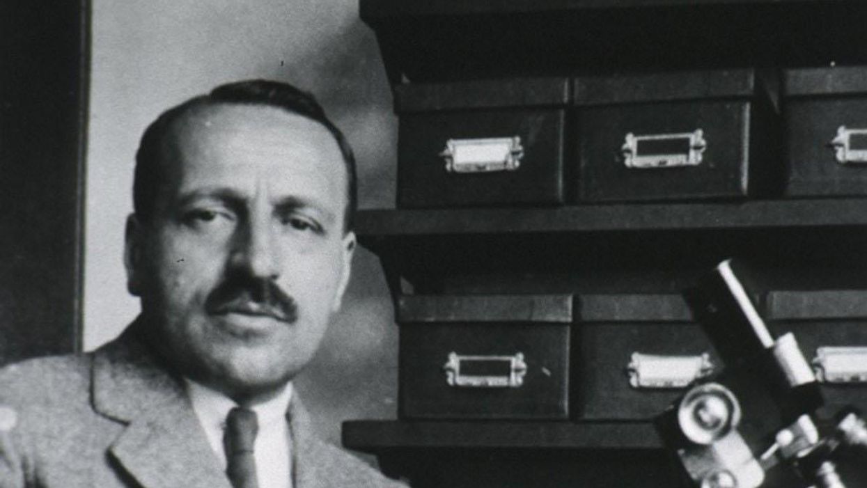
George Papanicolaou (1883-1962), Greek-born American physician developed a simple cytological test for cervical cancer in 1928.
For decades, women around the world have made the annual pilgrimage to their doctor for the dreaded but potentially life-saving Papanicolaou test, a gynecological exam to screen for cervical cancer named for Georgios Papanicolaou, the Greek immigrant who developed it.
The Pap smear, as it is commonly known, is credited for reducing cervical cancer mortality by 70% since the 1960s; the American Cancer Society (ACS) still ranks the Pap as the most successful screening test for preventing serious malignancies. Nonetheless, the agency, as well as other medical panels, including the US Preventive Services Task Force and the American College of Obstetrics and Gynecology are making a strong push to replace the Pap with the more sensitive high-risk HPV screening test for the human papillomavirus virus, which causes nearly all cases of cervical cancer.
So, how was the Pap developed and how did it become the gold standard of cervical cancer detection for more than 60 years?
Born on May 13, 1883, on the island of Euboea, Greece, Georgios Papanicolaou attended the University of Athens where he majored in music and the humanities before earning his medical degree in 1904 and PhD from the University of Munich six years later. In Europe, Papanicolaou was an assistant military surgeon during the Balkan War, a psychologist for an expedition of the Oceanographic Institute of Monaco and a caregiver for leprosy patients.
When he and his wife, Andromache Mavroyenous (Mary), arrived at Ellis Island on October 19, 1913, the young couple had scarcely more than the $250 minimum required to immigrate, spoke no English and had no job prospects. They worked a series of menial jobs--department store sales clerk, rug salesman, newspaper clerk, restaurant violinist--before Papanicolaou landed a position as an anatomy assistant at Cornell University and Mary was hired as his lab assistant, an arrangement that would last for the next 50 years.
Papanikolaou would later say the discovery "was one of the greatest thrills I ever experienced during my scientific career."
In his early research, Papanikolaou used guinea pigs to prove that gender is determined by the X and Y chromosomes. Using a pediatric nasal speculum, he collected and microscopically examined vaginal secretions of guinea pigs, which revealed distinct cell changes connected to the menstrual cycle. He moved on to study reproductive patterns in humans, beginning with his faithful wife, Mary, who not only endured his almost-daily cervical exams for decades, but also recruited friends as early research participants.
Writing in the medical journal Growth in 1920, the scientist outlined his theory that a microscopic smear of vaginal fluid could detect the presence of cancer cells in the uterus. Papanikolaou would later say the discovery "was one of the greatest thrills I ever experienced during my scientific career."
At this time, cervical cancer was the number one cancer killer of American women but physicians were skeptical of these new findings. They continued to rely on biopsy and curettage to diagnose and treat the disease until Papanicolaou's discovery was published in American Journal of Obstetrics and Gynecology. An inexpensive, easy-to-perform test that could detect cervical cancer, precancerous dysplasia and other cytological diseases was a sea change. Between 1975 and 2001, the cervical cancer rate was cut in half.
Papanicolaou became Emeritus Professor at Cornell University Medical College and received numerous awards, including the Albert Lasker Award for Clinical Medical Research and the Medal of Honor from the American Cancer Society. His image was featured on the Greek currency and the US Post Office issued a commemorative stamp in his honor. But international acclaim didn't lead to a more relaxed schedule. The researcher continued to work seven days a week and refused to take vacations.
After nearly 50 years, Papanicolaou left Cornell to head and develop the Cancer Institute of Miami. He died of a heart attack on February 19, 1962, just three months after his arrival. Mary continued to work in the renamed Papanicolaou Cancer Research Institute until her death 20 years later.
The annual pap smear was originally tied to renewing a birth control prescription. Canada began recommending Pap exams every three years in 1978. The United States followed suit in 2012, noting that it takes many years for cervical cancer to develop. In September 2020, the American Cancer Society recommended delaying the first gynecological pelvic exam until age 25 and replacing the Pap test completely with the more accurate human papillomavirus (HPV) test every five years as the technology becomes more widely available.
Not everyone agrees that it's time to do away with this proven screening method, though. The incidence rate of cervical cancer among Hispanic women is 28% higher than for white women, and Black women are more likely to die of cervical cancer than any other racial or ethnicities.
Whether the Pap is administered every year, every three years or not at all, Papanicolaou will always be known as the medical hero who saved countless women who would otherwise have succumbed to cervical cancer.

