The Dangers of Hype: How a Bold Claim and Sensational Media Unraveled a Company

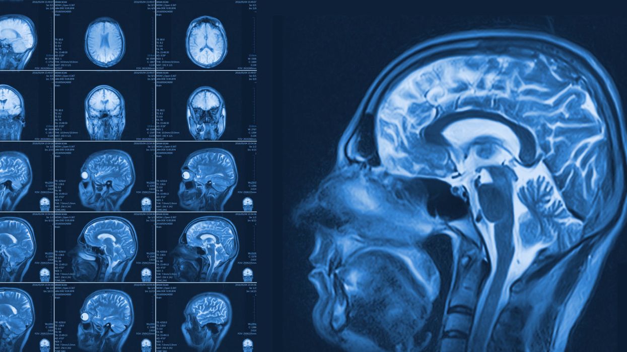
Magnetic resonance imaging of the brain.
This past March, headlines suddenly flooded the Internet about a startup company called Nectome. Founded by two graduates of the Massachusetts Institute of Technology, the new company was charging people $10,000 to join a waiting list to have their brains embalmed, down to the last neuron, using an award-winning chemical compound.
While the lay public presumably burnt their wills and grew ever more excited about the end of humanity's quest for immortality, neurologists let out a collective sigh.
Essentially, participants' brains would turn to a substance like glass and remain in a state of near-perfect preservation indefinitely. "If memories can truly be preserved by a sufficiently good brain banking technique," Nectome's website explains, "we believe that within the century it could become feasible to digitize your preserved brain and use that information to recreate your mind." But as with most Faustian bargains, Nectome's proposition came with a serious caveat -- death.
That's right, in order for Nectome's process to properly preserve your connectome, the comprehensive map of the brain's neural connections, you must be alive (and under anesthesia) while the fluid is injected. This way, the company postulates, when the science advances enough to read and extract your memories someday, your vitrified brain will still contain your perfectly preserved essence--which can then be digitally recreated as a computer simulation.
Almost immediately this story gained buzz with punchy headlines: "Startup wants to upload your brain to the cloud, but has to kill you to do it," "San Junipero is real: Nectome wants to upload your brain," and "New tech firm promises eternal life, but you have to die."
While the lay public presumably burnt their wills and grew ever more excited about the end of humanity's quest for immortality, neurologists let out a collective sigh -- hype had struck the scientific community once again.
The truth about Nectome is that its claims are highly speculative and no hard science exists to suggest that our connectome is the key to our 'being,' nor that it can ever be digitally revived. "We haven't come even close to understanding even the most basic types of functioning in the brain," says neuroscientist Alex Fox, who was educated at the University of Queensland in Australia. "Memory storage in the brain is only a theoretical concept [and] there are some seriously huge gaps in our knowledge base that stand in the way of testing [the connectome] theory."
After the Nectome story broke, Harvard computational neuroscientist Sam Gershman tweeted out:
"Didn't anyone tell them that we've known the C Elegans (a microscopic worm) connectome for over a decade but haven't figured out how to reconstruct all of their memories? And that's only 7000 synapses compared to the trillions of synapses in the human brain!"
Hype can come from researchers themselves, who are under an enormous amount of pressure to publish original work and maintain funding.
How media coverage of Nectome went from an initial fastidiously researched article in the MIT Technology Review by veteran science journalist Antonio Regalado to the click-bait frenzy it became is a prime example of the 'science hype' phenomenon. According to Adam Auch, who holds a doctorate in philosophy from Dalhousie University in Nova Scotia, Canada, "Hype is a feature of all stages of the scientific dissemination process, from the initial circulation of preliminary findings within particular communities of scientists, to the process by which such findings come to be published in peer-reviewed journals, to the subsequent uptake these findings receive from the non-specialist press and the general public."
In the case of Nectome, hype was present from the word go. Riding the high of several major wins, including having raised over one million dollars in funding and partnering with well-known MIT neurologist Edward Boyden, Nectome founders Michael McCanna and Robert McIntyre launched their website on March 1, 2018. Just one month prior, they were able to purchase and preserve a newly deceased corpse in Portland, Oregon, showing that vitrifixation, their method of chemical preservation, could be used on a human specimen. It had previously won an award for preserving every synaptic structure on a rabbit brain.
The Nectome mission statement, found on its website, is laced with saccharine language that skirts the unproven nature of the procedure the company is peddling for big bucks: "Our mission is to preserve your brain well enough to keep all its memories intact: from that great chapter of your favorite book to the feeling of cold winter air, baking an apple pie, or having dinner with your friends and family."
This rhetoric is an example of hype that can come from researchers themselves, who are under an enormous amount of pressure to publish original work and maintain funding. As a result, there is a constant push to present science as "groundbreaking" when really, as is apparently the case with Nectome, it is only a small piece in a much larger effort.
Calling out the audacity of Nectome's posited future, neuroscientist Gershman commented to another publication, "The important question is whether the connectome is sufficient for memory: Can I reconstruct all memories knowing only the connections between neurons? The answer is almost certainly no, given our knowledge about how memories are stored (itself a controversial topic)."

The former home page of Nectome's website, which has now been replaced by a statement titled, "Response to recent press."
Furthermore, universities like MIT, who entered into a subcontract with Nectome, are under pressure to seek funding through partnerships with industry as a result of the Bayh-Dole Act of 1980. Also known as the Patent and Trademark Law Amendments Act, this piece of legislation allows universities to commercialize inventions developed under federally funded research programs, like Nectome's method of preserving brains, formally called Aldehyde-Stabilized Cryopreservation.
"[Universities use] every incentive now to talk about innovation," explains Dr. Ivan Oransky, president of the Association of Health Care Journalists and co-founder of retractionwatch.com, a blog that catalogues errors and fraud in published research. "Innovation to me is often a fancy word for hype. The role of journalists should not be to glorify what universities [say, but to] tell the closest version of the truth they can."
In this case, a combination of the hyperbolic press, combined with some impressively researched expose pieces, led MIT to cut its ties with Nectome on April 2nd, 2018, just two weeks after the news of their company broke.
The solution to the dangers of hype, experts say, is a more scientifically literate public—and less clickbait-driven journalism.
Because of its multi-layered nature, science hype carries several disturbing consequences. For one, exaggerated coverage of a discovery could mislead the public by giving them false hope or unfounded worry. And media hype can contribute to a general mistrust of science. In these instances, people might, as Auch puts it, "fall back on previously held beliefs, evocative narratives, or comforting biases instead of well-justified scientific evidence."
All of this is especially dangerous in today's 'fake news' era, when companies or political parties sow public confusion for their own benefit, such as with global warming. In the case of Nectome, the danger is that people might opt to end their lives based off a lacking scientific theory. In fact, the company is hoping to enlist terminal patients in California, where doctor-assisted suicide is legal. And 25 people have paid the $10,000 to join Nectome's waiting list, including Sam Altman, president of the famed startup accelerator Y Combinator. Nectome now has offered to refund the money.
Founders McCanna and McIntyre did not return repeated requests for comment for this article. A new statement on their website begins: "Vitrifixation today is a powerful research tool, but needs more research and development before anyone considers applying it in a context other than research."
The solution to the dangers of hype, experts say, is a more scientifically literate public—and less clickbait-driven journalism. Until then, it seems that companies like Nectome will continue to enjoy at least 15 minutes of fame.
A new type of cancer therapy is shrinking deadly brain tumors with just one treatment
MRI scans after a new kind of immunotherapy for brain cancer show remarkable progress in one patient just days after the first treatment.
Few cancers are deadlier than glioblastomas—aggressive and lethal tumors that originate in the brain or spinal cord. Five years after diagnosis, less than five percent of glioblastoma patients are still alive—and more often, glioblastoma patients live just 14 months on average after receiving a diagnosis.
But an ongoing clinical trial at Mass General Cancer Center is giving new hope to glioblastoma patients and their families. The trial, called INCIPIENT, is meant to evaluate the effects of a special type of immune cell, called CAR-T cells, on patients with recurrent glioblastoma.
How CAR-T cell therapy works
CAR-T cell therapy is a type of cancer treatment called immunotherapy, where doctors modify a patient’s own immune system specifically to find and destroy cancer cells. In CAR-T cell therapy, doctors extract the patient’s T-cells, which are immune system cells that help fight off disease—particularly cancer. These T-cells are harvested from the patient and then genetically modified in a lab to produce proteins on their surface called chimeric antigen receptors (thus becoming CAR-T cells), which makes them able to bind to a specific protein on the patient’s cancer cells. Once modified, these CAR-T cells are grown in the lab for several weeks so that they can multiply into an army of millions. When enough cells have been grown, these super-charged T-cells are infused back into the patient where they can then seek out cancer cells, bind to them, and destroy them. CAR-T cell therapies have been approved by the US Food and Drug Administration (FDA) to treat certain types of lymphomas and leukemias, as well as multiple myeloma, but haven’t been approved to treat glioblastomas—yet.
CAR-T cell therapies don’t always work against solid tumors, such as glioblastomas. Because solid tumors contain different kinds of cancer cells, some cells can evade the immune system’s detection even after CAR-T cell therapy, according to a press release from Massachusetts General Hospital. For the INCIPIENT trial, researchers modified the CAR-T cells even further in hopes of making them more effective against solid tumors. These second-generation CAR-T cells (called CARv3-TEAM-E T cells) contain special antibodies that attack EFGR, a protein expressed in the majority of glioblastoma tumors. Unlike other CAR-T cell therapies, these particular CAR-T cells were designed to be directly injected into the patient’s brain.
The INCIPIENT trial results
The INCIPIENT trial involved three patients who were enrolled in the study between March and July 2023. All three patients—a 72-year-old man, a 74-year-old man, and a 57-year-old woman—were treated with chemo and radiation and enrolled in the trial with CAR-T cells after their glioblastoma tumors came back.
The results, which were published earlier this year in the New England Journal of Medicine (NEJM), were called “rapid” and “dramatic” by doctors involved in the trial. After just a single infusion of the CAR-T cells, each patient experienced a significant reduction in their tumor sizes. Just two days after receiving the infusion, the glioblastoma tumor of the 72-year-old man decreased by nearly twenty percent. Just two months later the tumor had shrunk by an astonishing 60 percent, and the change was maintained for more than six months. The most dramatic result was in the 57-year-old female patient, whose tumor shrank nearly completely after just one infusion of the CAR-T cells.
The results of the INCIPIENT trial were unexpected and astonishing—but unfortunately, they were also temporary. For all three patients, the tumors eventually began to grow back regardless of the CAR-T cell infusions. According to the press release from MGH, the medical team is now considering treating each patient with multiple infusions or prefacing each treatment with chemotherapy to prolong the response.
While there is still “more to do,” says co-author of the study neuro-oncologist Dr. Elizabeth Gerstner, the results are still promising. If nothing else, these second-generation CAR-T cell infusions may someday be able to give patients more time than traditional treatments would allow.
“These results are exciting but they are also just the beginning,” says Dr. Marcela Maus, a doctor and professor of medicine at Mass General who was involved in the clinical trial. “They tell us that we are on the right track in pursuing a therapy that has the potential to change the outlook for this intractable disease.”
A recent study in The Lancet Oncology showed that AI found 20 percent more cancers on mammogram screens than radiologists alone.
Since the early 2000s, AI systems have eliminated more than 1.7 million jobs, and that number will only increase as AI improves. Some research estimates that by 2025, AI will eliminate more than 85 million jobs.
But for all the talk about job security, AI is also proving to be a powerful tool in healthcare—specifically, cancer detection. One recently published study has shown that, remarkably, artificial intelligence was able to detect 20 percent more cancers in imaging scans than radiologists alone.
Published in The Lancet Oncology, the study analyzed the scans of 80,000 Swedish women with a moderate hereditary risk of breast cancer who had undergone a mammogram between April 2021 and July 2022. Half of these scans were read by AI and then a radiologist to double-check the findings. The second group of scans was read by two researchers without the help of AI. (Currently, the standard of care across Europe is to have two radiologists analyze a scan before diagnosing a patient with breast cancer.)
The study showed that the AI group detected cancer in 6 out of every 1,000 scans, while the radiologists detected cancer in 5 per 1,000 scans. In other words, AI found 20 percent more cancers than the highly-trained radiologists.

But even though the AI was better able to pinpoint cancer on an image, it doesn’t mean radiologists will soon be out of a job. Dr. Laura Heacock, a breast radiologist at NYU, said in an interview with CNN that radiologists do much more than simply screening mammograms, and that even well-trained technology can make errors. “These tools work best when paired with highly-trained radiologists who make the final call on your mammogram. Think of it as a tool like a stethoscope for a cardiologist.”
AI is still an emerging technology, but more and more doctors are using them to detect different cancers. For example, researchers at MIT have developed a program called MIRAI, which looks at patterns in patient mammograms across a series of scans and uses an algorithm to model a patient's risk of developing breast cancer over time. The program was "trained" with more than 200,000 breast imaging scans from Massachusetts General Hospital and has been tested on over 100,000 women in different hospitals across the world. According to MIT, MIRAI "has been shown to be more accurate in predicting the risk for developing breast cancer in the short term (over a 3-year period) compared to traditional tools." It has also been able to detect breast cancer up to five years before a patient receives a diagnosis.
The challenges for cancer-detecting AI tools now is not just accuracy. AI tools are also being challenged to perform consistently well across different ages, races, and breast density profiles, particularly given the increased risks that different women face. For example, Black women are 42 percent more likely than white women to die from breast cancer, despite having nearly the same rates of breast cancer as white women. Recently, an FDA-approved AI device for screening breast cancer has come under fire for wrongly detecting cancer in Black patients significantly more often than white patients.
As AI technology improves, radiologists will be able to accurately scan a more diverse set of patients at a larger volume than ever before, potentially saving more lives than ever.

