The Stunning Comeback of a Top Transplant Surgeon Who Got a New Heart at His Own Hospital

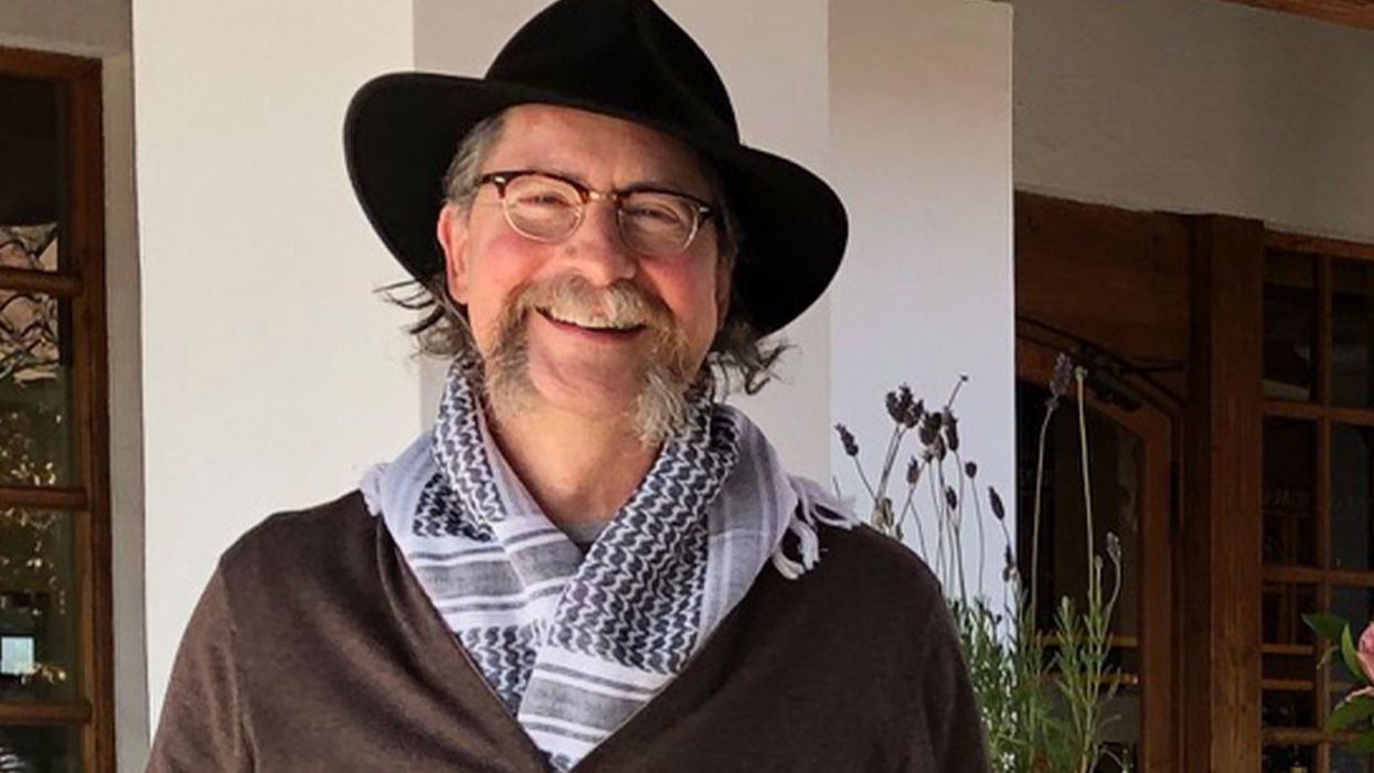
Dr. Robert Montgomery, almost one year post-transplant, at a vineyard in Casablanca, Chile, August 2019.
Having spent my working life as a transplant surgeon, it is the ultimate irony that I have now become a heart transplant patient. I knew this was a possibility since 1987, when I was 27 years old and I received a phone call from my sister-in-law telling me that my 35-year-old brother, Rich, had just died suddenly while water skiing.
Living from one heartbeat to the next I knew I had to get it right and nail my life—and in that regard my disease was a blessing.
After his autopsy, dots were connected and it was clear that the mysterious heart disease my father had died from when I was 15 years old was genetic. I was evaluated and it was clear that I too had inherited cardiomyopathy, a progressive weakening condition of the heart muscle that often leads to dangerous rhythm disturbances and sudden death. My doctors urged me to have a newly developed device called an implantable cardioverter-defibrillator (ICD) surgically placed in my abdomen and chest to monitor and shock my heart back into normal rhythm should I have a sudden cardiac arrest.
They also told me I was the first surgeon in the world to undergo an ICD implant and that having one of these devices would not be compatible with the life of a surgeon and I should change careers to something less rigorous. With the support of a mentor and armed with what the British refer to as my "bloody-mindedness," I refused to give up this dream of becoming a transplant surgeon. I completed my surgical training and embarked on my career.
What followed were periods of stability punctuated by near-death experiences. I had a family, was productive in my work, and got on with life, knowing that this was a fragile situation that could turn on its head in a moment. In a way, it made my decisions about how to spend my time and focus my efforts more deliberate and purposeful. Living from one heartbeat to the next I knew I had to get it right and nail my life—and in that regard my disease was a blessing.
In 2017 while pursuing my passion for the outdoors in a remote part of Patagonia, I collapsed from bacterial pneumonia and sepsis. Unknowingly, I had brought in my lungs one of those super-bugs that you read about from the hospital where I worked. Several days into the trip, the bacteria entered my blood stream and brought me as close to death as a human can get.
I lay for nearly 3 weeks in a coma on a stretcher in a tiny hospital in Argentina, septic and in cardiogenic shock before stabilizing enough to be evaced to NYU Langone Hospital, where I was on staff. I awoke helpless, unable to walk, talk, or swallow food or drink. It was a long shot but I managed to recover completely from this episode; after 3 months, I returned to work and the operating room. My heart rebounded, but never back to where it had been.
Then, on the eve of my mother's funeral, I arrested while watching a Broadway show, and this time my ICD failed to revive me. There was prolonged CPR that broke my ribs and spine and a final shock that recaptured my heart. It was literally a show stopper and I awoke to a standing ovation from the New York theatre audience who were stunned by my modern recreation of the biblical story of Lazarus, or for the more hip among them, my real-life rendition of the resurrection of Jon Snow at the end of season 5 of Game of Thrones.
Against the advice of my doctors, I attended my mom's funeral and again tried to regain some sense of normalcy. We discussed a transplant at this point but, believe it or not, there is such a scarcity of organs I was not yet "sick enough" to get enough priority to receive a heart. I had more surgery to supercharge my ICD so it would be more likely to save my life the next time -- and there would be a next time, I knew.
As a transplant surgeon, I have been involved in some important innovations to expand the number of organs available for transplantation.
Months later in Matera, Italy, where I was attending a medical meeting, I developed what is referred to as ventricular tachycardia storm. I had 4 cardiac arrests over a 3-hour period. With the first one, I fell on to a stone floor and split my forehead open. When I arrived at the small hospital it seemed like Patagonia all over again. One of the first people I met was a Catholic priest who gave me the Last Rights.
I knew now was the moment and so with the help of one of my colleagues who was at the meeting with me and the compassion of the Italian doctors who supplied my friend with resuscitation medications and left my IV in place, I signed out of the hospital against medical advice and boarded a commercial flight back to New York. I was admitted to the NYU intensive care unit and received a heart transplant 3 weeks later.
Now, what I haven't said is that as a transplant surgeon, I have been involved in some important innovations to expand the number of organs available for transplantation. I came to NYU in 2016 to start a new Transplant Institute which included inaugurating a heart transplant program. We hired heart transplant surgeons, cardiologists, and put together a team that unbeknownst to me at the time, would save my life a year later.
It gets even more interesting. One of the innovations that I had been involved in from its inception in the 1990s was using organs from donors at risk for transmitting viruses like HIV and Hepatitis C (Hep C). We popularized new ways to detect these viruses in donors and ensure that the risk was minimized as much as possible so patients in need of a life-saving transplant could utilize these organs.
When the opioid crisis hit hard about four years ago, there were suddenly a lot of potential donors who were IV drug users and 25 percent of them were known to be infected with Hep C (which is spread by needles). In 2018, 49,000 people died in the U.S. from drug overdoses. There were many more donors with Hep C than potential recipients who had previously been exposed to Hep C, and so more than half of these otherwise perfectly good organs were being discarded. At the same time, a new class of drugs was being tested that could cure Hep C.
I was at Johns Hopkins at the time and our team developed a protocol for using these Hep C positive organs for Hep C negative recipients who were willing to take them, even knowing that they were likely to become infected with the virus. We would then treat them after the transplant with this new class of drugs and in all likelihood, cure them. I brought this protocol with me to NYU.
When my own time came, I accepted a Hep C heart from a donor who overdosed on heroin. I became infected with Hep C and it was then eliminated from my body with 2 months of anti-viral therapy. All along this unlikely journey, I was seemingly making decisions that would converge upon that moment in time when I would arise to catch the heart that was meant for me.

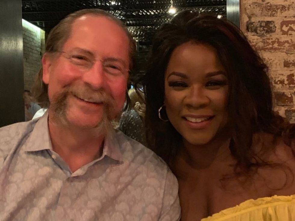
Dr. Montgomery with his wife Denyce Graves, September 2019.
(Courtesy Montgomery)
Today, I am almost exactly one year post-transplant, back to work, operating, traveling, enjoying the outdoors, and giving lectures. My heart disease is gone; gone when my heart was removed. Gone also is my ICD. I am no longer at risk for a sudden cardiac death. I traded all that for the life of a transplant patient, which has its own set of challenges, but I clearly traded up. It is cliché, I know, but I enjoy every moment of every day. It is a miracle I am still here.
A new type of cancer therapy is shrinking deadly brain tumors with just one treatment
MRI scans after a new kind of immunotherapy for brain cancer show remarkable progress in one patient just days after the first treatment.
Few cancers are deadlier than glioblastomas—aggressive and lethal tumors that originate in the brain or spinal cord. Five years after diagnosis, less than five percent of glioblastoma patients are still alive—and more often, glioblastoma patients live just 14 months on average after receiving a diagnosis.
But an ongoing clinical trial at Mass General Cancer Center is giving new hope to glioblastoma patients and their families. The trial, called INCIPIENT, is meant to evaluate the effects of a special type of immune cell, called CAR-T cells, on patients with recurrent glioblastoma.
How CAR-T cell therapy works
CAR-T cell therapy is a type of cancer treatment called immunotherapy, where doctors modify a patient’s own immune system specifically to find and destroy cancer cells. In CAR-T cell therapy, doctors extract the patient’s T-cells, which are immune system cells that help fight off disease—particularly cancer. These T-cells are harvested from the patient and then genetically modified in a lab to produce proteins on their surface called chimeric antigen receptors (thus becoming CAR-T cells), which makes them able to bind to a specific protein on the patient’s cancer cells. Once modified, these CAR-T cells are grown in the lab for several weeks so that they can multiply into an army of millions. When enough cells have been grown, these super-charged T-cells are infused back into the patient where they can then seek out cancer cells, bind to them, and destroy them. CAR-T cell therapies have been approved by the US Food and Drug Administration (FDA) to treat certain types of lymphomas and leukemias, as well as multiple myeloma, but haven’t been approved to treat glioblastomas—yet.
CAR-T cell therapies don’t always work against solid tumors, such as glioblastomas. Because solid tumors contain different kinds of cancer cells, some cells can evade the immune system’s detection even after CAR-T cell therapy, according to a press release from Massachusetts General Hospital. For the INCIPIENT trial, researchers modified the CAR-T cells even further in hopes of making them more effective against solid tumors. These second-generation CAR-T cells (called CARv3-TEAM-E T cells) contain special antibodies that attack EFGR, a protein expressed in the majority of glioblastoma tumors. Unlike other CAR-T cell therapies, these particular CAR-T cells were designed to be directly injected into the patient’s brain.
The INCIPIENT trial results
The INCIPIENT trial involved three patients who were enrolled in the study between March and July 2023. All three patients—a 72-year-old man, a 74-year-old man, and a 57-year-old woman—were treated with chemo and radiation and enrolled in the trial with CAR-T cells after their glioblastoma tumors came back.
The results, which were published earlier this year in the New England Journal of Medicine (NEJM), were called “rapid” and “dramatic” by doctors involved in the trial. After just a single infusion of the CAR-T cells, each patient experienced a significant reduction in their tumor sizes. Just two days after receiving the infusion, the glioblastoma tumor of the 72-year-old man decreased by nearly twenty percent. Just two months later the tumor had shrunk by an astonishing 60 percent, and the change was maintained for more than six months. The most dramatic result was in the 57-year-old female patient, whose tumor shrank nearly completely after just one infusion of the CAR-T cells.
The results of the INCIPIENT trial were unexpected and astonishing—but unfortunately, they were also temporary. For all three patients, the tumors eventually began to grow back regardless of the CAR-T cell infusions. According to the press release from MGH, the medical team is now considering treating each patient with multiple infusions or prefacing each treatment with chemotherapy to prolong the response.
While there is still “more to do,” says co-author of the study neuro-oncologist Dr. Elizabeth Gerstner, the results are still promising. If nothing else, these second-generation CAR-T cell infusions may someday be able to give patients more time than traditional treatments would allow.
“These results are exciting but they are also just the beginning,” says Dr. Marcela Maus, a doctor and professor of medicine at Mass General who was involved in the clinical trial. “They tell us that we are on the right track in pursuing a therapy that has the potential to change the outlook for this intractable disease.”
A recent study in The Lancet Oncology showed that AI found 20 percent more cancers on mammogram screens than radiologists alone.
Since the early 2000s, AI systems have eliminated more than 1.7 million jobs, and that number will only increase as AI improves. Some research estimates that by 2025, AI will eliminate more than 85 million jobs.
But for all the talk about job security, AI is also proving to be a powerful tool in healthcare—specifically, cancer detection. One recently published study has shown that, remarkably, artificial intelligence was able to detect 20 percent more cancers in imaging scans than radiologists alone.
Published in The Lancet Oncology, the study analyzed the scans of 80,000 Swedish women with a moderate hereditary risk of breast cancer who had undergone a mammogram between April 2021 and July 2022. Half of these scans were read by AI and then a radiologist to double-check the findings. The second group of scans was read by two researchers without the help of AI. (Currently, the standard of care across Europe is to have two radiologists analyze a scan before diagnosing a patient with breast cancer.)
The study showed that the AI group detected cancer in 6 out of every 1,000 scans, while the radiologists detected cancer in 5 per 1,000 scans. In other words, AI found 20 percent more cancers than the highly-trained radiologists.

But even though the AI was better able to pinpoint cancer on an image, it doesn’t mean radiologists will soon be out of a job. Dr. Laura Heacock, a breast radiologist at NYU, said in an interview with CNN that radiologists do much more than simply screening mammograms, and that even well-trained technology can make errors. “These tools work best when paired with highly-trained radiologists who make the final call on your mammogram. Think of it as a tool like a stethoscope for a cardiologist.”
AI is still an emerging technology, but more and more doctors are using them to detect different cancers. For example, researchers at MIT have developed a program called MIRAI, which looks at patterns in patient mammograms across a series of scans and uses an algorithm to model a patient's risk of developing breast cancer over time. The program was "trained" with more than 200,000 breast imaging scans from Massachusetts General Hospital and has been tested on over 100,000 women in different hospitals across the world. According to MIT, MIRAI "has been shown to be more accurate in predicting the risk for developing breast cancer in the short term (over a 3-year period) compared to traditional tools." It has also been able to detect breast cancer up to five years before a patient receives a diagnosis.
The challenges for cancer-detecting AI tools now is not just accuracy. AI tools are also being challenged to perform consistently well across different ages, races, and breast density profiles, particularly given the increased risks that different women face. For example, Black women are 42 percent more likely than white women to die from breast cancer, despite having nearly the same rates of breast cancer as white women. Recently, an FDA-approved AI device for screening breast cancer has come under fire for wrongly detecting cancer in Black patients significantly more often than white patients.
As AI technology improves, radiologists will be able to accurately scan a more diverse set of patients at a larger volume than ever before, potentially saving more lives than ever.

