Small changes in how a person talks could reveal Alzheimer’s earlier

In an initial study, Canary analyzed speech recordings with AI and identified early stage Alzheimer’s with 96 percent accuracy.
Dave Arnold retired in his 60s and began spending time volunteering in local schools. But then he started misplacing items, forgetting appointments and losing his sense of direction. Eventually he was diagnosed with early stage Alzheimer’s.
“Hearing the diagnosis made me very emotional and tearful,” he said. “I immediately thought of all my mom had experienced.” His mother suffered with the condition for years before passing away. Over the last year, Arnold has worked for the Alzheimer’s Association as one of its early stage advisors, sharing his insights to help others in the initial stages of the disease.
Arnold was diagnosed sooner than many others. It's important to find out early, when interventions can make the most difference. One promising avenue is looking at how people talk. Research has shown that Alzheimer’s affects a part of the brain that controls speech, resulting in small changes before people show other signs of the disease.
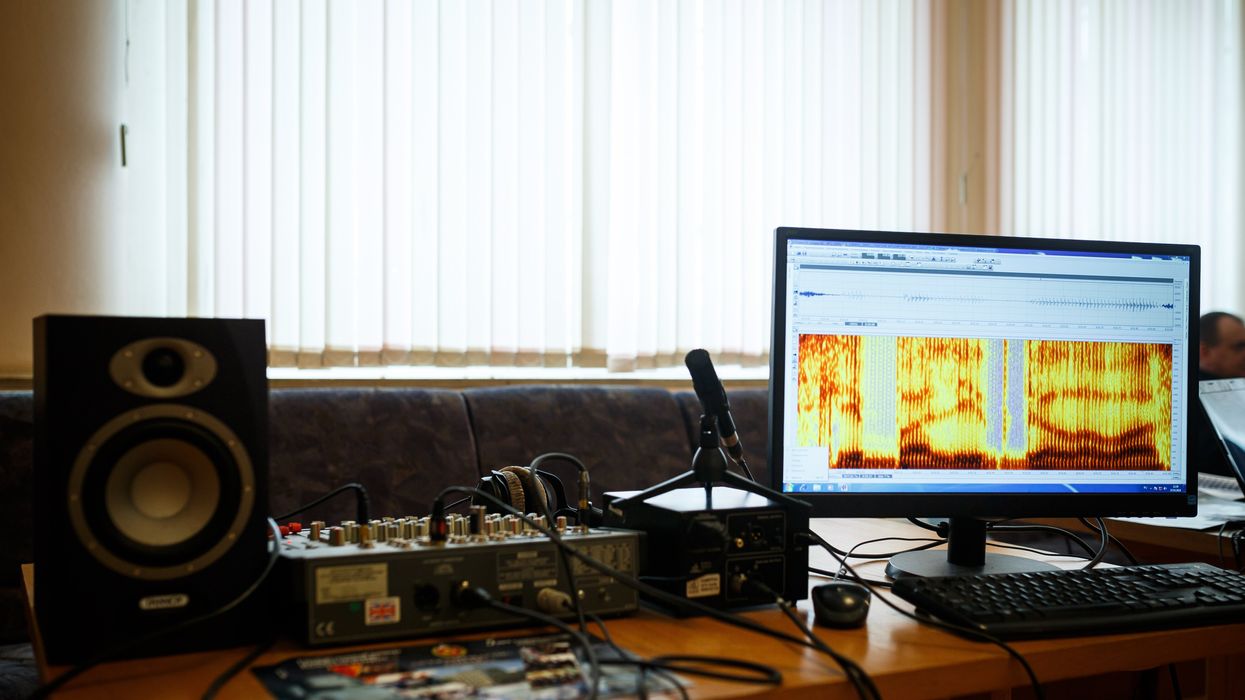
Now, Canary Speech, a company based in Utah, is using AI to examine elements like the pitch of a person’s voice and their pauses. In an initial study, Canary analyzed speech recordings with AI and identified early stage Alzheimer’s with 96 percent accuracy.
Developing the AI model
Canary Speech’s CEO, Henry O’Connell, met cofounder Jeff Adams about 40 years before they started the company. Back when they first crossed paths, they were both living in Bethesda, Maryland; O’Connell was a research fellow at the National Institutes of Health studying rare neurological diseases, while Adams was working to decode spy messages. Later on, Adams would specialize in building mathematical models to analyze speech and sound as a team leader in developing Amazon's Alexa.
It wasn't until 2015 that they decided to make use of the fit between their backgrounds. ““We established Canary Speech in 2017 to build a product that could be used in multiple languages in clinical environments,” O'Connell says.
The need is growing. About 55 million people worldwide currently live with Alzheimer’s, a number that is expected to double by 2050. Some scientists think the disease results from a buildup of plaque in the brain. It causes mild memory loss at first and, over time, this issue get worse while other symptoms, such as disorientation and hallucinations, can develop. Treatment to manage the disease is more effective in the earlier stages, but detection is difficult since mild symptoms are often attributed to the normal aging process.
O’Connell and Adams specialize in the complex ways that Alzheimer’s effects how people speak. Using AI, their mathematical model analyzes 15 million data points every minute, focusing on certain features of speech such as pitch, pauses and elongation of words. It also pays attention to how the vibrations of vocal cords change in different stages of the disease.
To create their model, the team used a type of machine learning called deep neural nets, which looks at multiple layers of data - in this case, the multiple features of a person’s speech patterns.
“Deep neural nets allow us to look at much, much larger data sets built out of millions of elements,” O’Connell explained. “Through machine learning and AI, we’ve identified features that are very sensitive to an Alzheimer’s patient versus [people without the disease] and also very sensitive to mild cognitive impairment, early stage and moderate Alzheimer's.” Based on their learnings, Canary is able to classify the disease stage very quickly, O’Connell said.
“When we’re listening to sublanguage elements, we’re really analyzing the direct result of changes in the brain in the physical body,” O’Connell said. “The brain controls your vocal cords: how fast they vibrate, the expansion of them, the contraction.” These factors, along with where people put their tongues when talking, function subconsciously and result in subtle changes in the sounds of speech.
Further testing is needed
In an initial trial, Canary analyzed speech recordings from phone calls to a large U.S. health insurer. They looked at the audio recordings of 651 policyholders who had early stage Alzheimer’s and 1018 who did not have the condition, aiming for a representative sample of age, gender and race. They used this data to create their first diagnostic model and found that it was 96 percent accurate in identifying Alzheimer’s.
Christian Herff, an assistant professor of neuroscience at Maastricht University in the Netherlands, praised this approach while adding that further testing is needed to assess its effectiveness.
“I think the general idea of identifying increased risk for cognitive impairment based on speech characteristics is very feasible, particularly when change in a user’s voice is monitored, for example, by recording speech every year,” Herff said. He noted that this can only be a first indication, not a full diagnosis. The accuracy still needs to be validated in studies that follows individuals over a period of time, he said.
Toby Walsh, a professor of artificial intelligence at the University of New South Wales, also thinks Canary’s tool has potential but highlights that Canary could diagnose some people who don’t really have the disease. “This is an interesting and promising application of AI,” he said, “but these tools need to be used carefully. Imagine the anxiety of being misdiagnosed with Alzheimer’s.”
As with many other AI tools, privacy and bias are additional issues to monitor closely, Walsh said.
Other languages
A related issue is that not everyone is fluent in English. Mahnaz Arvaneh, a senior lecturer in automatic control and systems engineering at the University of Sheffield, said this could be a blind spot.
“The system may not be very accurate for those who have English as their second language as their speaking patterns would be different, and any issue might be because of language deficiency rather than cognitive issues,” Arvaneh said.
The team is expanding to multiple languages starting with Japanese and Spanish. The elements of the model that make up the algorithm are very similar, but they need to be validated and retrained in a different language, which will require access to more data.
Recently, Canary analyzed the phone calls of 233 Japanese patients who had mild cognitive impairment and 704 healthy people. Using an English model they were able to identify the Japanese patients who had mild cognitive impairment with 78 percent accuracy. They also developed a model in Japanese that was 45 percent accurate, and they’re continuing to train it with more data.
The future
Canary is using their model to look at other diseases like Huntington’s and Parkinson’s. They’re also collaborating with pharmaceuticals to validate potential therapies for Alzheimer’s. By looking at speech patterns over time, Canary can get an indication of how well these drugs are working.

Dave Arnold and his wife dance at his nephew’s wedding in Rochester, New York, ten years ago, before his Alzheimer's diagnosis.
Dave Arnold
Ultimately, they want to integrate their tool into everyday life. “We want it to be used in a smartphone, or a teleconference call so that individuals could be examined in their home,” O’Connell said. “We could follow them over time and work with clinical teams and hospitals to improve the evaluation of patients and contribute towards an accurate diagnosis.”
Arnold, the patient with early stage Alzheimer’s, sees great promise. “The process of getting a diagnosis is already filled with so much anxiety,” he said. “Anything that can be done to make it easier and less stressful would be a good thing, as long as it’s proven accurate.”
A new type of cancer therapy is shrinking deadly brain tumors with just one treatment
MRI scans after a new kind of immunotherapy for brain cancer show remarkable progress in one patient just days after the first treatment.
Few cancers are deadlier than glioblastomas—aggressive and lethal tumors that originate in the brain or spinal cord. Five years after diagnosis, less than five percent of glioblastoma patients are still alive—and more often, glioblastoma patients live just 14 months on average after receiving a diagnosis.
But an ongoing clinical trial at Mass General Cancer Center is giving new hope to glioblastoma patients and their families. The trial, called INCIPIENT, is meant to evaluate the effects of a special type of immune cell, called CAR-T cells, on patients with recurrent glioblastoma.
How CAR-T cell therapy works
CAR-T cell therapy is a type of cancer treatment called immunotherapy, where doctors modify a patient’s own immune system specifically to find and destroy cancer cells. In CAR-T cell therapy, doctors extract the patient’s T-cells, which are immune system cells that help fight off disease—particularly cancer. These T-cells are harvested from the patient and then genetically modified in a lab to produce proteins on their surface called chimeric antigen receptors (thus becoming CAR-T cells), which makes them able to bind to a specific protein on the patient’s cancer cells. Once modified, these CAR-T cells are grown in the lab for several weeks so that they can multiply into an army of millions. When enough cells have been grown, these super-charged T-cells are infused back into the patient where they can then seek out cancer cells, bind to them, and destroy them. CAR-T cell therapies have been approved by the US Food and Drug Administration (FDA) to treat certain types of lymphomas and leukemias, as well as multiple myeloma, but haven’t been approved to treat glioblastomas—yet.
CAR-T cell therapies don’t always work against solid tumors, such as glioblastomas. Because solid tumors contain different kinds of cancer cells, some cells can evade the immune system’s detection even after CAR-T cell therapy, according to a press release from Massachusetts General Hospital. For the INCIPIENT trial, researchers modified the CAR-T cells even further in hopes of making them more effective against solid tumors. These second-generation CAR-T cells (called CARv3-TEAM-E T cells) contain special antibodies that attack EFGR, a protein expressed in the majority of glioblastoma tumors. Unlike other CAR-T cell therapies, these particular CAR-T cells were designed to be directly injected into the patient’s brain.
The INCIPIENT trial results
The INCIPIENT trial involved three patients who were enrolled in the study between March and July 2023. All three patients—a 72-year-old man, a 74-year-old man, and a 57-year-old woman—were treated with chemo and radiation and enrolled in the trial with CAR-T cells after their glioblastoma tumors came back.
The results, which were published earlier this year in the New England Journal of Medicine (NEJM), were called “rapid” and “dramatic” by doctors involved in the trial. After just a single infusion of the CAR-T cells, each patient experienced a significant reduction in their tumor sizes. Just two days after receiving the infusion, the glioblastoma tumor of the 72-year-old man decreased by nearly twenty percent. Just two months later the tumor had shrunk by an astonishing 60 percent, and the change was maintained for more than six months. The most dramatic result was in the 57-year-old female patient, whose tumor shrank nearly completely after just one infusion of the CAR-T cells.
The results of the INCIPIENT trial were unexpected and astonishing—but unfortunately, they were also temporary. For all three patients, the tumors eventually began to grow back regardless of the CAR-T cell infusions. According to the press release from MGH, the medical team is now considering treating each patient with multiple infusions or prefacing each treatment with chemotherapy to prolong the response.
While there is still “more to do,” says co-author of the study neuro-oncologist Dr. Elizabeth Gerstner, the results are still promising. If nothing else, these second-generation CAR-T cell infusions may someday be able to give patients more time than traditional treatments would allow.
“These results are exciting but they are also just the beginning,” says Dr. Marcela Maus, a doctor and professor of medicine at Mass General who was involved in the clinical trial. “They tell us that we are on the right track in pursuing a therapy that has the potential to change the outlook for this intractable disease.”
A recent study in The Lancet Oncology showed that AI found 20 percent more cancers on mammogram screens than radiologists alone.
Since the early 2000s, AI systems have eliminated more than 1.7 million jobs, and that number will only increase as AI improves. Some research estimates that by 2025, AI will eliminate more than 85 million jobs.
But for all the talk about job security, AI is also proving to be a powerful tool in healthcare—specifically, cancer detection. One recently published study has shown that, remarkably, artificial intelligence was able to detect 20 percent more cancers in imaging scans than radiologists alone.
Published in The Lancet Oncology, the study analyzed the scans of 80,000 Swedish women with a moderate hereditary risk of breast cancer who had undergone a mammogram between April 2021 and July 2022. Half of these scans were read by AI and then a radiologist to double-check the findings. The second group of scans was read by two researchers without the help of AI. (Currently, the standard of care across Europe is to have two radiologists analyze a scan before diagnosing a patient with breast cancer.)
The study showed that the AI group detected cancer in 6 out of every 1,000 scans, while the radiologists detected cancer in 5 per 1,000 scans. In other words, AI found 20 percent more cancers than the highly-trained radiologists.

But even though the AI was better able to pinpoint cancer on an image, it doesn’t mean radiologists will soon be out of a job. Dr. Laura Heacock, a breast radiologist at NYU, said in an interview with CNN that radiologists do much more than simply screening mammograms, and that even well-trained technology can make errors. “These tools work best when paired with highly-trained radiologists who make the final call on your mammogram. Think of it as a tool like a stethoscope for a cardiologist.”
AI is still an emerging technology, but more and more doctors are using them to detect different cancers. For example, researchers at MIT have developed a program called MIRAI, which looks at patterns in patient mammograms across a series of scans and uses an algorithm to model a patient's risk of developing breast cancer over time. The program was "trained" with more than 200,000 breast imaging scans from Massachusetts General Hospital and has been tested on over 100,000 women in different hospitals across the world. According to MIT, MIRAI "has been shown to be more accurate in predicting the risk for developing breast cancer in the short term (over a 3-year period) compared to traditional tools." It has also been able to detect breast cancer up to five years before a patient receives a diagnosis.
The challenges for cancer-detecting AI tools now is not just accuracy. AI tools are also being challenged to perform consistently well across different ages, races, and breast density profiles, particularly given the increased risks that different women face. For example, Black women are 42 percent more likely than white women to die from breast cancer, despite having nearly the same rates of breast cancer as white women. Recently, an FDA-approved AI device for screening breast cancer has come under fire for wrongly detecting cancer in Black patients significantly more often than white patients.
As AI technology improves, radiologists will be able to accurately scan a more diverse set of patients at a larger volume than ever before, potentially saving more lives than ever.

