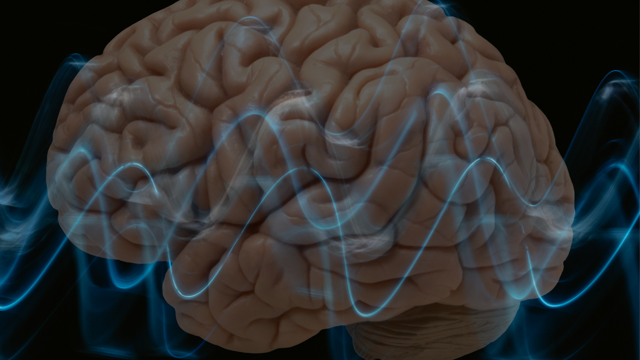
Scientists Are Harnessing Sound Waves in Hopes of Treating Alzheimer’s

Researchers at Columbia University are testing an experimental treatment for Alzheimer's that uses ultrasound waves and "microbubbles."
In 2010, a 67-year-old former executive assistant for a Fortune 500 company was diagnosed with mild cognitive impairment. By 2014, her doctors confirmed she had Alzheimer's disease.
As her disease progressed, she continued to live independently but wasn't able to drive anymore. Today, she can manage most of her everyday tasks, but her two daughters are considering a live-in caregiver. Despite her condition, the woman may represent a beacon of hope for the approximately 44 million people worldwide living with Alzheimer's disease. The now 74-year-old is among a small cadre of Alzheimer's patients who have undergone an experimental ultrasound procedure aimed at slowing cognitive decline.
In November 2020, Elisa Konofagou, a professor of biomedical engineering and director of the Ultrasound and Elasticity Imaging Laboratory at Columbia University, and her team used ultrasound to noninvasively open the woman's blood-brain barrier. This barrier is a highly selective membrane of cells that prevents toxins and pathogens from entering the brain while allowing vital nutrients to pass through. This regulatory function means the blood-brain barrier filters out most drugs, making treating Alzheimer's and other brain diseases a challenge.
Ultrasound uses high-frequency sound waves to produce live images from the inside of the human body. But scientists think it could also be used to boost the effectiveness of Alzheimer's drugs, or potentially even improve brain function in dementia patients without the use of drugs.
The procedure, which involves a portable ultrasound system, is the culmination of 17 years of lab work. As part of a small clinical trial, scientists positioned a sensor transmitting ultrasound waves on top of the woman's head while she sat in a chair. The sensor sends ultrasound pulses throughout the target region. Meanwhile, investigators intravenously infused microbubbles into the woman to boost the effects of the ultrasound. Three days after the procedure, scientists scanned her brain so that they could measure the effects of the treatments. Five months later, they took more images of her brain to see if the effects of the treatment lasted.
Promising Signs
After the first brain scan, Konofagou and her team found that amyloid-beta, the protein that clumps together in the brains of Alzheimer's patients and disrupts cell function, had declined by 14%. At the woman's second scan, amyloid levels were still lower than before the experimental treatment, but only by 10% this time. Konofagou thinks repeat ultrasound treatments given early on in the development of Alzheimer's may have the best chance at keeping amyloid plaques at bay.
This reduction in amyloid appeared to halt the woman's cognitive decline, at least temporarily. Following the ultrasound treatment, the woman took a 30-point test used to measure cognitive impairment in Alzheimer's. Her score — 22, indicating mild cognitive impairment — remained the same as before the intervention. Konofagou says this was actually a good sign.
"Typically, every six months an Alzheimer's patient scores two to three points lower, so this is highly encouraging," she says.
Konofagou speculates that the results might have been even more impressive had they applied the ultrasound on a larger section of the brain at a higher frequency. The selected site was just 4 cubic centimeters. Current safety protocols set by the U.S. Food and Drug Administration stipulate that investigators conducting such trials only treat one brain region with the lowest pressure possible.
The Columbia trial is aided by microbubble technology. During the procedure, investigators infused tiny, gas-filled spheres into the woman's veins to enhance the ultrasound reflection of the sound waves.
The big promise of ultrasound is that it could eventually make drugs for Alzheimer's obsolete.
"Ultrasound with microbubbles wakes up immune cells that go on to discard amyloid-beta," Konofagou says. "In this way, we can recover the function of brain neurons, which are destroyed by Alzheimer's in a sort of domino effect." What's more, a drug delivered alongside ultrasound can penetrate the brain at a dose up to 10 times higher.
Costas Arvanitis, an assistant professor at Georgia Institute of Technology who studies ultrasonic biophysics and isn't involved in the Columbia trial, is excited about the research. "First, by applying ultrasound you can make larger drugs — picture an antibody — available to the brain," he says. Then, you can use ultrasound to improve the therapeutic index, or the ratio of the effectiveness of a drug versus the ratio of adverse effects. "Some drugs might be effective but because we have to provide them in high doses to see significant responses they tend to come with side effects. By improving locally the concentration of a drug, you open up the possibility to reduce the dose."
The Columbia trial will enroll just six patients and is designed to test the feasibility and safety of the approach, not its efficacy. Still, Arvantis is hopeful about the potential benefits of the technique. "The technology has already been demonstrated to be safe, its components are now tuned to the needs of this specific application, and it's safe to say it's only a matter of time before we are able to develop personalized treatments," he says.
Konofagou and her colleagues recently presented their findings at the 20th Annual International Symposium for Therapeutic Ultrasound and intend to publish them in a scientific journal later this year. They plan to recruit more participants for larger trials, which will determine how effective the therapy is at improving memory and brain function in Alzheimer's patients. They're also in talks with pharmaceutical companies about ways to use their therapeutic approach to improve current drugs or even "create new drugs," says Konofagou.
A New Treatment Approach
On June 7, the FDA approved the first Alzheimer's disease drug in nearly two decades. Aducanumab, a drug developed by Biogen, is an antibody designed to target and reduce amyloid plaques. The drug has already sparked immense enthusiasm — and controversy. Proponents say the drug is a much-needed start in the fight against the disease, but others argue that the drug doesn't substantially improve cognition. They say the approval could open the door to the FDA greenlighting more Alzheimer's drugs that don't have a clear benefit, giving false hope to both patients and their families.
Konofagou's ultrasound approach could potentially boost the effects of drugs like aducanumab. "Our technique can be seamlessly combined with aducanumab in early Alzheimer's, where it has shown the most promise, to further enhance both its amyloid load reduction and further reduce cognitive deficits while using exactly the same drug regimen otherwise," she says. For the Columbia team, the goal is to use ultrasound to maximize the effects of aducanumab, as they've done with other drugs in animal studies.
But Konofagou's approach could transcend drug controversies, and even drugs altogether. The big promise of ultrasound is that it could eventually make drugs for Alzheimer's obsolete.
"There are already indications that the immune system is alerted each time ultrasound is exerted on the brain or when the brain barrier is being penetrated and gets activated, which on its own may have sufficient therapeutic effects," says Konofagou. Her team is now working with psychiatrists in hopes of using brain stimulation to treat patients with depression.
The potential to modulate the brain without drugs is huge and untapped, says Kim Butts Pauly, a professor of radiology, electrical engineering and bioengineering at Stanford University, who's not involved in the Columbia study. But she admits that scientists don't know how to fully control ultrasound in the brain yet. "We're only at the starting point of getting the tools to understand and harness how ultrasound microbubbles stimulate an immune response in the brain."
Meanwhile, the 74-year-old woman who received the ultrasound treatment last year, goes on about her life, having "both good days and bad days," her youngest daughter says. COVID-19's isolation took a toll on her, but both she and her daughters remain grateful for the opportunity to participate in the ultrasound trial.
"My mother wants to help, if not for herself, then for those who will follow her," the daughter says. She hopes her mother will be able to join the next phase of the trial, which will involve a drug in conjunction with the ultrasound treatment. "This may be the combination where the magic will happen," her daughter says.
Indigenous wisdom plus honeypot ants could provide new antibiotics
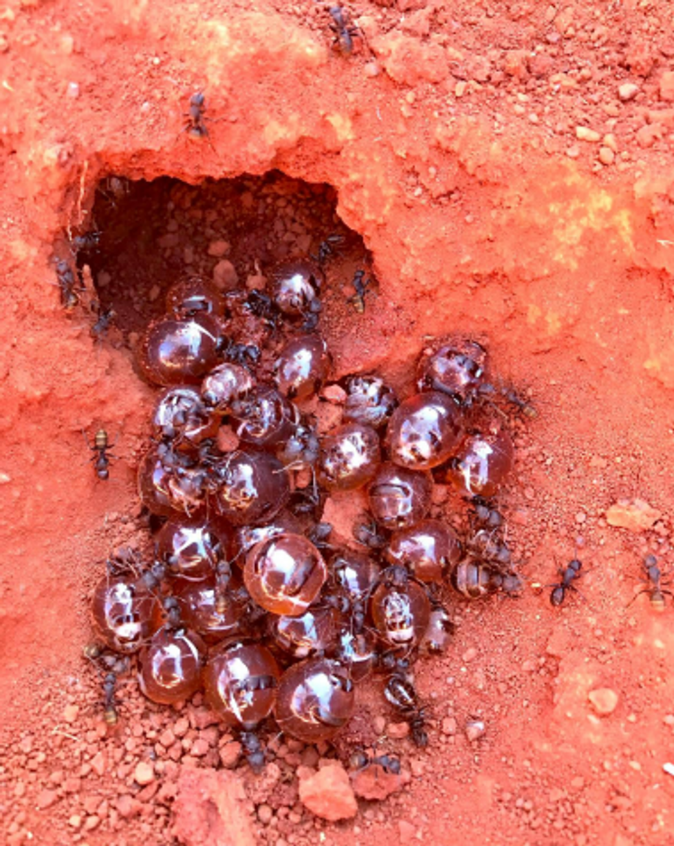
Indigenous people in Australia dig pits next to a honeypot colony. Scientists think the honey can be used to make new antimicrobial drugs.
For generations, the Indigenous Tjupan people of Australia enjoyed the sweet treat of honey made by honeypot ants. As a favorite pastime, entire families would go searching for the underground colonies, first spotting a worker ant and then tracing it to its home. The ants, which belong to the species called Camponotus inflatus, usually build their subterranean homes near the mulga trees, Acacia aneura. Having traced an ant to its tree, it would be the women who carefully dug a pit next to a colony, cautious not to destroy the entire structure. Once the ant chambers were exposed, the women would harvest a small amount to avoid devastating the colony’s stocks—and the family would share the treat.
The Tjupan people also knew that the honey had antimicrobial properties. “You could use it for a sore throat,” says Danny Ulrich, a member of the Tjupan nation. “You could also use it topically, on cuts and things like that.”
These hunts have become rarer, as many of the Tjupan people have moved away and, up until now, the exact antimicrobial properties of the ant honey remained unknown. But recently, scientists Andrew Dong and Kenya Fernandes from the University of Sydney, joined Ulrich, who runs the Honeypot Ants tours in Kalgoorlie, a city in Western Australia, on a honey-gathering expedition. Afterwards, they ran a series of experiments analyzing the honey’s antimicrobial activity—and confirmed that the Indigenous wisdom was true. The honey was effective against Staphylococcus aureus, a common pathogen responsible for sore throats, skin infections like boils and sores, and also sepsis, which can result in death. Moreover, the honey also worked against two species of fungi, Cryptococcus and Aspergillus, which can be pathogenic to humans, especially those with suppressed immune systems.
In the era of growing antibiotic resistance and the rising threat of pathogenic fungi, these findings may help scientists identify and make new antimicrobial compounds. “Natural products have been honed over thousands and millions of years by nature and evolution,” says Fernandes. “And some of them have complex and intricate properties that make them really important as potential new antibiotics. “

In an era of growing resistance to antibiotics and new threats of fungi infections, the latest findings about honeypot ants are helping scientists identify new antimicrobial drugs.
Danny Ulrich
Bee honey is also known for its antimicrobial properties, but bees produce it very differently than the ants. Bees collect nectar from flowers, which they regurgitate at the hive and pack into the hexagonal honeycombs they build for storage. As they do so, they also add into the mix an enzyme called glucose oxidase produced by their glands. The enzyme converts atmospheric oxygen into hydrogen peroxide, a reactive molecule that destroys bacteria and acts as a natural preservative. After the bees pack the honey into the honeycombs, they fan it with their wings to evaporate the water. Once a honeycomb is full, the bees put a beeswax cover on it, where it stays well-preserved thanks to the enzymatic action, until the bees need it.
Less is known about the chemistry of ants’ honey-making. Similarly to bees, they collect nectar. They also collect the sweet sap of the mulga tree. Additionally, they also “milk” the aphids—small sap-sucking insects that live on the tree. When ants tickle the aphids with their antennae, the latter release a sweet substance, which the former also transfer to their colonies. That’s where the honey management difference becomes really pronounced. The ants don’t build any kind of structures to store their honey. Instead, they store it in themselves.
The workers feed their harvest to their fellow ants called repletes, stuffing them up to the point that their swollen bellies outgrow the ants themselves, looking like amber-colored honeypots—hence the name. Because of their size, repletes don’t move, but hang down from the chamber’s ceiling, acting as living feedstocks. When food becomes scarce, they regurgitate their reserves to their colony’s brethren. It’s not clear whether the repletes die afterwards or can be restuffed again. “That's a good question,” Dong says. “After they've been stretched, they can't really return to exactly the same shape.”
These replete ants are the “treat” the Tjupan women dug for. Once they saw the round-belly ants inside the chambers, they would reach in carefully and get a few scoops of them. “You see a lot of honeypot ants just hanging on the roof of the little openings,” says Ulrich’s mother, Edie Ulrich. The women would share the ants with family members who would eat them one by one. “They're very delicate,” shares Edie Ulrich—you have to take them out carefully, so they don’t accidentally pop and become a wasted resource. “Because you’d lose all this precious honey.”
Dong stumbled upon the honeypot ants phenomenon because he was interested in Indigenous foods and went on Ulrich’s tour. He quickly became fascinated with the insects and their role in the Indigenous culture. “The honeypot ants are culturally revered by the Indigenous people,” he says. Eventually he decided to test out the honey’s medicinal qualities.
The researchers were surprised to see that even the smallest, eight percent concentration of honey was able to arrest the growth of S. aureus.
To do this, the two scientists first diluted the ant honey with water. “We used something called doubling dilutions, which means that we made 32 percent dilutions, and then we halve that to 16 percent and then we half that to eight percent,” explains Fernandes. The goal was to obtain as much results as possible with the meager honey they had. “We had very, very little of the honeypot ant honey so we wanted to maximize the spectrum of results we can get without wasting too much of the sample.”
After that, the researchers grew different microbes inside a nutrient rich broth. They added the broth to the different honey dilutions and incubated the mixes for a day or two at the temperature favorable to the germs’ growth. If the resulting solution turned turbid, it was a sign that the bugs proliferated. If it stayed clear, it meant that the honey destroyed them. The researchers were surprised to see that even the smallest, eight percent concentration of honey was able to arrest the growth of S. aureus. “It was really quite amazing,” Fernandes says. “Eight milliliters of honey in 92 milliliters of water is a really tiny amount of honey compared to the amount of water.”
Similar to bee honey, the ants’ honey exhibited some peroxide antimicrobial activity, researchers found, but given how little peroxide was in the solution, they think the honey also kills germs by a different mechanism. “When we measured, we found that [the solution] did have some hydrogen peroxide, but it didn't have as much of it as we would expect based on how active it was,” Fernandes says. “Whether this hydrogen peroxide also comes from glucose oxidase or whether it's produced by another source, we don't really know,” she adds. The research team does have some hypotheses about the identity of this other germ-killing agent. “We think it is most likely some kind of antimicrobial peptide that is actually coming from the ant itself.”
The honey also has a very strong activity against the two types of fungi, Cryptococcus and Aspergillus. Both fungi are associated with trees and decaying leaves, as well as in the soils where ants live, so the insects likely have evolved some natural defense compounds, which end up inside the honey.
It wouldn’t be the first time when modern medicines take their origin from the natural world or from the indigenous people’s knowledge. The bark of the cinchona tree native to South America contains quinine, a substance that treats malaria. The Indigenous people of the Andes used the bark to quell fever and chills for generations, and when Europeans began to fall ill with malaria in the Amazon rainforest, they learned to use that medicine from the Andean people.
The wonder drug aspirin similarly takes its origin from a bark of a tree—in this case a willow.
Even some anticancer compounds originated from nature. A chemotherapy drug called Paclitaxel, was originally extracted from the Pacific yew trees, Taxus brevifolia. The samples of the Pacific yew bark were first collected in 1962 by researchers from the United States Department of Agriculture who were looking for natural compounds that might have anti-tumor activity. In December 1992, the FDA approved Paclitaxel (brand name Taxol) for the treatment of ovarian cancer and two years later for breast cancer.
In the era when the world is struggling to find new medicines fast enough to subvert a fungal or bacterial pandemic, these discoveries can pave the way to new therapeutics. “I think it's really important to listen to indigenous cultures and to take their knowledge because they have been using these sources for a really, really long time,” Fernandes says. Now we know it works, so science can elucidate the molecular mechanisms behind it, she adds. “And maybe it can even provide a lead for us to develop some kind of new treatments in the future.”
Lina Zeldovich has written about science, medicine and technology for Popular Science, Smithsonian, National Geographic, Scientific American, Reader’s Digest, the New York Times and other major national and international publications. A Columbia J-School alumna, she has won several awards for her stories, including the ASJA Crisis Coverage Award for Covid reporting, and has been a contributing editor at Nautilus Magazine. In 2021, Zeldovich released her first book, The Other Dark Matter, published by the University of Chicago Press, about the science and business of turning waste into wealth and health. You can find her on http://linazeldovich.com/ and @linazeldovich.
Blood Test Can Detect Lymphoma Cells Before a Tumor Grows Back
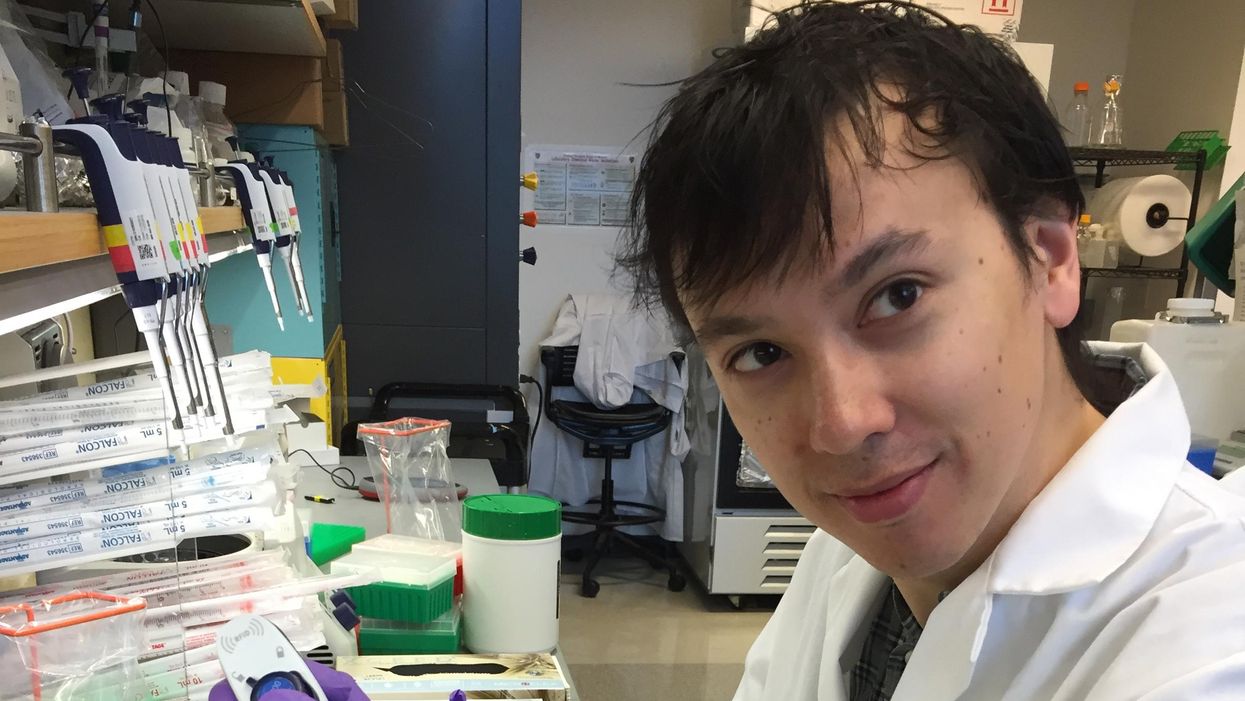
David Kurtz making DNA sequencing libraries in his lab.
When David M. Kurtz was doing his clinical fellowship at Stanford University Medical Center in 2009, specializing in lymphoma treatments, he found himself grappling with a question no one could answer. A typical regimen for these blood cancers prescribed six cycles of chemotherapy, but no one knew why. "The number seemed to be drawn out of a hat," Kurtz says. Some patients felt much better after just two doses, but had to endure the toxic effects of the entire course. For some elderly patients, the side effects of chemo are so harsh, they alone can kill. Others appeared to be cancer-free on the CT scans after the requisite six but then succumbed to it months later.
"Anecdotally, one patient decided to stop therapy after one dose because he felt it was so toxic that he opted for hospice instead," says Kurtz, now an oncologist at the center. "Five years down the road, he was alive and well. For him, just one dose was enough." Others would return for their one-year check up and find that their tumors grew back. Kurtz felt that while CT scans and MRIs were powerful tools, they weren't perfect ones. They couldn't tell him if there were any cancer cells left, stealthily waiting to germinate again. The scans only showed the tumor once it was back.
Blood cancers claim about 68,000 people a year, with a new diagnosis made about every three minutes, according to the Leukemia Research Foundation. For patients with B-cell lymphoma, which Kurtz focuses on, the survival chances are better than for some others. About 60 percent are cured, but the remaining 40 percent will relapse—possibly because they will have a negative CT scan, but still harbor malignant cells. "You can't see this on imaging," says Michael Green, who also treats blood cancers at University of Texas MD Anderson Medical Center.
The new blood test is sensitive enough to spot one cancerous perpetrator amongst one million other DNA molecules.
Kurtz wanted a better diagnostic tool, so he started working on a blood test that could capture the circulating tumor DNA or ctDNA. For that, he needed to identify the specific mutations typical for B-cell lymphomas. Working together with another fellow PhD student Jake Chabon, Kurtz finally zeroed-in on the tumor's genetic "appearance" in 2017—a pair of specific mutations sitting in close proximity to each other—a rare and telling sign. The human genome contains about 3 billion base pairs of nucleotides—molecules that compose genes—and in case of the B-cell lymphoma cells these two mutations were only a few base pairs apart. "That was the moment when the light bulb went on," Kurtz says.
The duo formed a company named Foresight Diagnostics, focusing on taking the blood test to the clinic. But knowing the tumor's mutational signature was only half the process. The other was fishing the tumor's DNA out of patients' bloodstream that contains millions of other DNA molecules, explains Chabon, now Foresight's CEO. It would be like looking for an escaped criminal in a large crowd. Kurtz and Chabon solved the problem by taking the tumor's "mug shot" first. Doctors would take the biopsy pre-treatment and sequence the tumor, as if taking the criminal's photo. After treatments, they would match the "mug shot" to all DNA molecules derived from the patient's blood sample to see if any molecular criminals managed to escape the chemo.
Foresight isn't the only company working on blood-based tumor detection tests, which are dubbed liquid biopsies—other companies such as Natera or ArcherDx developed their own. But in a recent study, the Foresight team showed that their method is significantly more sensitive in "fishing out" the cancer molecules than existing tests. Chabon says that this test can detect circulating tumor DNA in concentrations that are nearly 100 times lower than other methods. Put another way, it's sensitive enough to spot one cancerous perpetrator amongst one million other DNA molecules.
They also aim to extend their test to detect other malignancies such as lung, breast or colorectal cancers.
"It increases the sensitivity of detection and really catches most patients who are going to progress," says Green, the University of Texas oncologist who wasn't involved in the study, but is familiar with the method. It would also allow monitoring patients during treatment and making better-informed decisions about which therapy regimens would be most effective. "It's a minimally invasive test," Green says, and "it gives you a very high confidence about what's going on."
Having shown that the test works well, Kurtz and Chabon are planning a new trial in which oncologists would rely on their method to decide when to stop or continue chemo. They also aim to extend their test to detect other malignancies such as lung, breast or colorectal cancers. The latest genome sequencing technologies have sequenced and catalogued over 2,500 different tumor specimens and the Foresight team is analyzing this data, says Chabon, which gives the team the opportunity to create more molecular "mug shots."
The team hopes that that their blood cancer test will become available to patients within about five years, making doctors' job easier, and not only at the biological level. "When I tell patients, "good news, your cancer is in remission', they ask me, 'does it mean I'm cured?'" Kurtz says. "Right now I can't answer this question because I don't know—but I would like to." His company's test, he hopes, will enable him to reply with certainty. He'd very much like to have the power of that foresight.
This article is republished from our archives to coincide with Blood Cancer Awareness Month, which highlights progress in cancer diagnostics and treatment.
Lina Zeldovich has written about science, medicine and technology for Popular Science, Smithsonian, National Geographic, Scientific American, Reader’s Digest, the New York Times and other major national and international publications. A Columbia J-School alumna, she has won several awards for her stories, including the ASJA Crisis Coverage Award for Covid reporting, and has been a contributing editor at Nautilus Magazine. In 2021, Zeldovich released her first book, The Other Dark Matter, published by the University of Chicago Press, about the science and business of turning waste into wealth and health. You can find her on http://linazeldovich.com/ and @linazeldovich.

