A recent study by researchers at the University of Pennsylvania examined how CAR-T therapy helped Doug Olson beat a cancer death sentence for over a decade - and how it could work for more people.
Doug Olson was 49 when he was diagnosed with chronic lymphocytic leukemia, a blood cancer that strikes 21,000 Americans annually. Although the disease kills most patients within a decade, Olson’s case progressed more slowly, and courses of mild chemotherapy kept him healthy for 13 years. Then, when he was 62, the medication stopped working. The cancer had mutated, his doctor explained, becoming resistant to standard remedies. Harsher forms of chemo might buy him a few months, but their side effects would be debilitating. It was time to consider the treatment of last resort: a bone-marrow transplant.
Olson, a scientist who developed blood-testing instruments, knew the odds. There was only a 50 percent chance that a transplant would cure him. There was a 20 percent chance that the agonizing procedure—which involves destroying the patient’s marrow with chemo and radiation, then infusing his blood with donated stem cells—would kill him. If he survived, he would face the danger of graft-versus-host disease, in which the donor’s cells attack the recipient’s tissues. To prevent it, he would have to take immunosuppressant drugs, increasing the risk of infections. He could end up with pneumonia if one of his three grandchildren caught a sniffle. “I was being pushed into a corner,” Olson recalls, “with very little room to move.”
Soon afterward, however, his doctor revealed a possible escape route. He and some colleagues at the University of Pennsylvania’s Abramson Cancer Center were starting a clinical trial, he said, and Olson—still mostly symptom-free—might be a good candidate. The experimental treatment, known as CAR-T therapy, would use genetic engineering to turn his T lymphocytes (immune cells that guard against viruses and other pathogens) into a weapon against cancer.
In September 2010, technicians took some of Olson’s T cells to a laboratory, where they were programmed with new molecular marching orders and coaxed to multiply into an army of millions. When they were ready, a nurse inserted a catheter into his neck. At the turn of a valve, his soldiers returned home, ready to do battle.
“I felt like I’d won the lottery,” Olson says. But he was only the second person in the world to receive this “living drug,” as the University of Pennsylvania investigators called it. No one knew how long his remission would last.
Three weeks later, Olson was slammed with a 102-degree fever, nausea, and chills. The treatment had triggered two dangerous complications: cytokine release syndrome, in which immune chemicals inflame the patient’s tissues, and tumor lysis syndrome, in which toxins from dying cancer cells overwhelm the kidneys. But the crisis passed quickly, and the CAR-T cells fought on. A month after the infusion, the doctor delivered astounding news: “We can’t find any cancer in your body.”
“I felt like I’d won the lottery,” Olson says. But he was only the second person in the world to receive this “living drug,” as the University of Pennsylvania investigators called it. No one knew how long his remission would last.
An Unexpected Cure
In February 2022, the same cancer researchers reported a remarkable milestone: the trial’s first two patients had survived for more than a decade. Although Olson’s predecessor—a retired corrections officer named Bill Ludwig—died of COVID-19 complications in early 2021, both men had remained cancer-free. And the modified immune cells continued to patrol their territory, ready to kill suspected tumor cells the moment they arose.
“We can now conclude that CAR-T cells can actually cure patients with leukemia,” University of Pennsylvania immunologist Carl June, who spearheaded the development of the technique, told reporters. “We thought the cells would be gone in a month or two. The fact that they’ve survived 10 years is a major surprise.”
Even before the announcement, it was clear that CAR-T therapy could win a lasting reprieve for many patients with cancers that were once a death sentence. Since the Food and Drug Administration approved June’s version (marketed as Kymriah) in 2017, the agency has greenlighted five more such treatments for various types of leukemia, lymphoma, and myeloma. “Every single day, I take care of patients who would previously have been told they had no options,” says Rayne Rouce, a pediatric hematologist/oncologist at Texas Children’s Cancer Center. “Now we not only have a treatment option for those patients, but one that could potentially be the last therapy for their cancer that they’ll ever have to receive.”

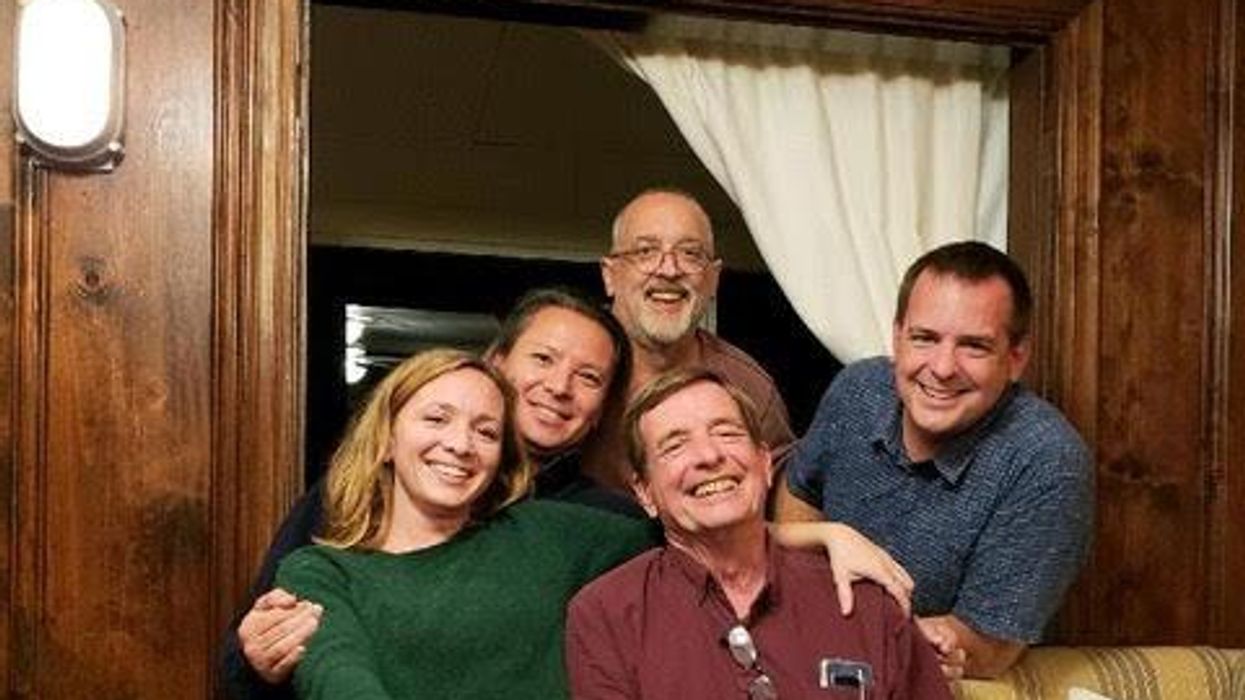
Immunologist Carl June, middle, spearheaded development of the CAR-T therapy that gave patients Bill Ludwig, left, and Doug Olson, right, a lengthy reprieve on their terminal cancer diagnoses.
Penn Medicine
Yet the CAR-T approach doesn’t help everyone. So far, it has only shown success for blood cancers—and for those, the overall remission rate is 30 to 40 percent. “When it works, it works extraordinarily well,” says Olson’s former doctor, David Porter, director of Penn’s blood and bone marrow transplant program. “It’s important to know why it works, but it’s equally important to know why it doesn’t—and how we can fix that.”
The team’s study, published in the journal Nature, offers a wealth of data on what worked for these two patients. It may also hold clues for how to make the therapy effective for more people.
Building a Better T Cell
Carl June didn’t set out to cure cancer, but his serendipitous career path—and a personal tragedy—helped him achieve insights that had eluded other researchers. In 1971, hoping to avoid combat in Vietnam, he applied to the U.S. Naval Academy in Annapolis, Maryland. June showed a knack for biology, so the Navy sent him on to Baylor College of Medicine. He fell in love with immunology during a fellowship researching malaria vaccines in Switzerland. Later, the Navy deployed him to the Fred Hutchinson Cancer Research Center in Seattle to study bone marrow transplantation.
There, June became part of the first research team to learn how to culture T cells efficiently in a lab. After moving on to the National Naval Medical Center in the ’80s, he used that knowledge to combat the newly emerging AIDS epidemic. HIV, the virus that causes the disease, invades T cells and eventually destroys them. June and his post-doc Bruce Levine developed a method to restore patients’ depleted cell populations, using tiny magnetic beads to deliver growth-stimulating proteins. Infused into the body, the new T cells effectively boosted immune function.
In 1999, after leaving the Navy, June joined the University of Pennsylvania. His wife, who’d been diagnosed with ovarian cancer, died two years later, leaving three young children. “I had not known what it was like to be on the other side of the bed,” he recalls. Watching her suffer through grueling but futile chemotherapy, followed by an unsuccessful bone-marrow transplant, he resolved to focus on finding better cancer treatments. He started with leukemia—a family of diseases in which mutant white blood cells proliferate in the marrow.
Cancer is highly skilled at slipping through the immune system’s defenses. T cells, for example, detect pathogens by latching onto them with receptors designed to recognize foreign proteins. Leukemia cells evade detection, in part, by masquerading as normal white blood cells—that is, as part of the immune system itself.
June planned to use a viral vector no one had tried before: HIV.
To June, chimeric antigen receptor (CAR) T cells looked like a promising tool for unmasking and destroying the impostors. Developed in the early ’90s, these cells could be programmed to identify a target protein, and to kill any pathogen that displayed it. To do the programming, you spliced together snippets of DNA and inserted them into a disabled virus. Next, you removed some of the patient’s T cells and infected them with the virus, which genetically hijacked its new hosts—instructing them to find and slay the patient’s particular type of cancer cells. When the T cells multiplied, their descendants carried the new genetic code. You then infused those modified cells into the patient, where they went to war against their designated enemy.
Or that’s what happened in theory. Many scientists had tried to develop therapies using CAR-T cells, but none had succeeded. Although the technique worked in lab animals, the cells either died out or lost their potency in humans.
But June had the advantage of his years nurturing T cells for AIDS patients, as well as the technology he’d developed with Levine (who’d followed him to Penn with other team members). He also planned to use a viral vector no one had tried before: HIV, which had evolved to thrive in human T cells and could be altered to avoid causing disease. By the summer of 2010, he was ready to test CAR-T therapy against chronic lymphocytic leukemia (CLL), the most common form of the disease in adults.
Three patients signed up for the trial, including Doug Olson and Bill Ludwig. A portion of each man’s T cells were reprogrammed to detect a protein found only on B lymphocytes, the type of white blood cells affected by CLL. Their genetic instructions ordered them to destroy any cell carrying the protein, known as CD19, and to multiply whenever they encountered one. This meant the patients would forfeit all their B cells, not just cancerous ones—but regular injections of gamma globulins (a cocktail of antibodies) would make up for the loss.
After being infused with the CAR-T cells, all three men suffered high fevers and potentially life-threatening inflammation, but all pulled through without lasting damage. The third patient experienced a partial remission and survived for eight months. Olson and Ludwig were cured.
Learning What Works
Since those first infusions, researchers have developed reliable ways to prevent or treat the side effects of CAR-T therapy, greatly reducing its risks. They’ve also been experimenting with combination therapies—pairing CAR-T with chemo, cancer vaccines, and immunotherapy drugs called checkpoint inhibitors—to improve its success rate. But CAR-T cells are still ineffective for at least 60 percent of blood cancer patients. And they remain in the experimental stage for solid tumors (including pancreatic cancer, mesothelioma, and glioblastoma), whose greater complexity make them harder to attack.
The new Nature study offers clues that could fuel further advances. The Penn team “profiled these cells at a level where we can almost say, ‘These are the characteristics that a T cell would need to survive 10 years,’” says Rouce, the physician at Texas Children’s Cancer Center.
One surprising finding involves how CAR-T cells change in the body over time. At first, those that Olson and Ludwig received showed the hallmarks of “killer” T-cells (also known as CD8 cells)—highly active lymphocytes bent on exterminating every tumor cell in sight. After several months, however, the population shifted toward “helper” T-cells (or CD4s), which aid in forming long-term immune memory but are normally incapable of direct aggression. Over the years, the numbers swung back and forth, until only helper cells remained. Those cells showed markers suggesting they were too exhausted to function—but in the lab, they were able not only to recognize but to destroy cancer cells.
June and his team suspect that those tired-looking helper cells had enough oomph to kill off any B cells Olson and Ludwig made, keeping the pair’s cancers permanently at bay. If so, that could prompt new approaches to selecting cells for CAR-T therapy. Maybe starting with a mix of cell types—not only CD8s, but CD4s and other varieties—would work better than using CD8s alone. Or perhaps inducing changes in cell populations at different times would help.
Another potential avenue for improvement is starting with healthier cells. Evidence from this and other trials hints that patients whose T cells are more robust to begin with respond better when their cells are used in CAR-T therapy. The Penn team recently completed a clinical trial in which CLL patients were treated with ibrutinib—a drug that enhances T-cell function—before their CAR-T cells were manufactured. The response rate, says David Porter, was “very high,” with most patients remaining cancer-free a year after being infused with the souped-up cells.
Such approaches, he adds, are essential to achieving the next phase in CAR-T therapy: “Getting it to work not just in more people, but in everybody.”

Doug Olson enjoys nature - and having a future.
Penn Medicine
To grasp what that could mean, it helps to talk with Doug Olson, who’s now 75. In the years since his infusion, he has watched his four children forge careers, and his grandkids reach their teens. He has built a business and enjoyed the rewards of semi-retirement. He’s done volunteer and advocacy work for cancer patients, run half-marathons, sailed the Caribbean, and ridden his bike along the sun-dappled roads of Silicon Valley, his current home.
And in his spare moments, he has just sat there feeling grateful. “You don’t really appreciate the effect of having a lethal disease until it’s not there anymore,” he says. “The world looks different when you have a future.”
This article was first published on Leaps.org on March 24, 2022.
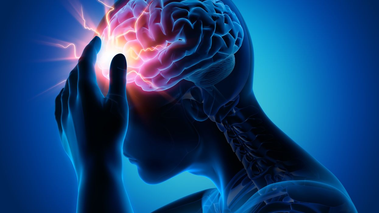
A new test called the EyeBox could provide a more objective - and portable - tool to measure whether people have concussions in stadiums and hospitals.
Concussion affects around 42 million people worldwide. While it’s increasingly common in the news because of sports injuries, anything that causes damage to the head, from a fall to a car accident, can result in a concussion. The sudden blow or jolt can disrupt the normal way the brain works. In the immediate aftermath, people may suffer from headaches, lose consciousness and experience dizziness, confusion and vomiting. Some recover but others have side effects that can last for years, particularly affecting memory and concentration.
There is no simple standard-of-care test to confirm a concussion or rule it out. Neither do they appear on MRI and CT scans. Instead, medical professionals use more indirect approaches that test symptoms of concussions, such as assessments of patients’ learning and memory skills, ability to concentrate and problem solving. They also look at balance and coordination. Most tests are in the form of questionnaires or symptom checklists. Consequently, they have limitations, can be biased and may miss a concussion or produce a false positive. Some people suspected of having a concussion may ordinarily have difficulties with literary and problem-solving tests because of language challenges or education levels.
Another problem with current tests is that patients, particularly soldiers who want to return to combat and athletes who would like to keep competing, could try and hide their symptoms to avoid being diagnosed with a brain injury. Trauma physicians who work with concussion patients have the need for a tool that is more objective and consistent.
“This type of assessment doesn’t rely on the patient's education level, willingness to follow instructions or cooperation. You can’t game this.” -- Uzma Samadani, founder of Oculogica
“The importance of having an objective measurement tool for the diagnosis of concussion is of great importance,” says Douglas Powell, associate professor of biomechanics at the University of Memphis, with research interests in sports injury and concussion. “While there are a number of promising systems or metrics, we have yet to develop a system that is portable, accessible and objective for use on the sideline and in the clinic. The EyeBOX may be able to address these issues, though time will be the ultimate test of performance.”
The EyeBOX as a window inside the brain
Using eye movements to diagnose a concussion has emerged as a promising technique since around 2010. Oculogica combined eye movements with AI to develop the EyeBOX to develop an unbiased objective diagnostic tool.
“What’s so great about this type of assessment is it doesn’t rely on the patient's education level, willingness to follow instructions or cooperation,” says Uzma Samadani, a neurosurgeon and brain injury researcher at the University of Minnesota, who founded Oculogica. “You can’t game this. It assesses functions that are prompted by your brain.”
In 2010, Samadani was working on a clinical trial to improve the outcome of brain injuries. The team needed some way to measure if seriously brain injured patients were improving. One thing patients could do was watch TV. So Samadani designed and patented an AI-based algorithm that tracks the relationship between eye movement and concussion.
The EyeBOX test requires patients to watch movie or music clips for 220 seconds. An eye tracking camera records subconscious eye movements, tracking eye positions 500 times per seconds as patients watch the video. It collects over 100,000 data points. The device then uses AI to assess whether there’s any disruptions from the normal way the eyes move.
Cranial nerves are responsible for transmitting information between the brain and the body. Many are involved in eye movement. Pressure caused by a concussion can affect how these nerves work. So tracking how the eyes move can indicate if there’s anything wrong with the cranial nerves and where the problem lies.
If someone is healthy, their eyes should be able to focus on an object, follow movement and both eyes should be coordinated with each other. The EyeBox can detect abnormalities. For example, if a patient’s eyes are coordinated but they are not moving as they should, that indicates issues in the central brain stem, whilst only one eye moving abnormally suggests that a particular nerve section is affected.
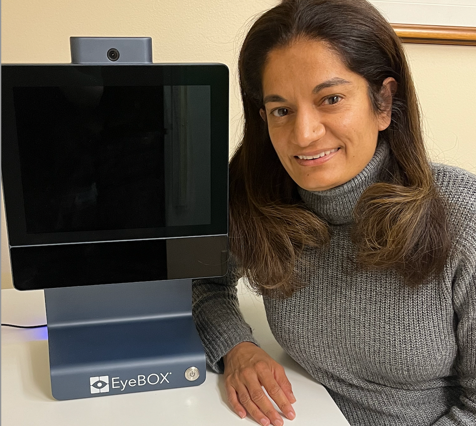
Uzma Samadani with the EyeBOX device
Courtesy Oculogica
“The EyeBOX is a monitor for cranial nerves,” says Samadani. “Essentially it’s a form of digital neurological exam. “Several other eye-tracking techniques already exist, but they rely on subjective self-reported symptoms. Many also require a baseline, a measure of how patients reacted when they were healthy, which often isn’t available.
VOMS (Vestibular Ocular Motor Screen) is one of the most accurate diagnostic tests used in clinics in combination with other tests, but it is subjective. It involves a therapist getting patients to move their head or eyes as they focus or follow a particular object. Patients then report their symptoms.
The King-Devick test measures how fast patients can read numbers and compares it to a baseline. Since it is mainly used for athletes, the initial test is completed before the season starts. But participants can manipulate it. It also cannot be used in emergency rooms because the majority of patients wouldn’t have prior baseline tests.
Unlike these tests, EyeBOX doesn’t use a baseline and is objective because it doesn’t rely on patients’ answers. “It shows great promise,” says Thomas Wilcockson, a senior lecturer of psychology in Loughborough University, who is an expert in using eye tracking techniques in neurological disorders. “Baseline testing of eye movements is not always possible. Alternative measures of concussion currently in development, including work with VR headsets, seem to currently require it. Therefore the EyeBOX may have an advantage.”
A technology that’s still evolving
In their last clinical trial, Oculogica used the EyeBOX to test 46 patients who had concussion and 236 patients who did not. The sensitivity of the EyeBOX, or the probability of it correctly identifying the patient’s concussion, was 80.4 percent. Meanwhile, the test accurately ruled out a concussion in 66.1 percent of cases. This is known as its specificity score.
While the team is working on improving the numbers, experts who treat concussion patients find the device promising. “I strongly support their use of eye tracking for diagnostic decision making,” says Douglas Powell. “But for diagnostic tests, we would prefer at least one of the sensitivity or specificity values to be greater than 90 percent. Powell compares EyeBOX with the Buffalo Concussion Treadmill Test, which has sensitivity and specificity values of 73 and 78 percent, respectively. The VOMS also has shown greater accuracy than the EyeBOX, at least for now. Still, EyeBOX is competitive with the best diagnostic testing available for concussion and Powell hopes that its detection prowess will improve. “I anticipate that the algorithms being used by Oculogica will be under continuous revision and expect the results will improve within the next several years.”
“The color of your skin can have a huge impact in how quickly you are triaged and managed for brain injury. People of color have significantly worse outcomes after traumatic brain injury than people who are white.” -- Uzma Samadani, founder of Oculogica
Powell thinks the EyeBOX could be an important complement to other concussion assessments.
“The Oculogica product is a viable diagnostic tool that supports clinical decision making. However, concussion is an injury that can present with a wide array of symptoms, and the use of technology such as the Oculogica should always be a supplement to patient interaction.”
Ioannis Mavroudis, a consultant neurologist at Leeds Teaching Hospital, agrees that the EyeBOX has promise, but cautions that concussions are too complex to rely on the device alone. For example, not all concussions affect how eyes move. “I believe that it can definitely help, however not all concussions show changes in eye movements. I believe that if this could be combined with a cognitive assessment the results would be impressive.”
The Oculogica team submitted their clinical data for FDA approval and received it in 2018. Now, they’re working to bring the test to the commercial market and using the device clinically to help diagnose concussions for clients. They also want to look at other areas of brain health in the next few years. Samadani believes that the EyeBOX could possibly be used to detect diseases like multiple sclerosis or other neurological conditions. “It’s a completely new way of figuring out what someone’s neurological exam is and we’re only beginning to realize the potential,” says Samadani.
One of Samadani’s biggest aspirations is to help reduce inequalities in healthcare because of skin color and other factors like money or language barriers. From that perspective, the EyeBOX’s greatest potential could be in emergency rooms. It can help diagnose concussions in addition to the questionnaires, assessments and symptom checklists, currently used in the emergency departments. Unlike these more subjective tests, EyeBOX can produce an objective analysis of brain injury through AI when patients are admitted and assessed, unrelated to their socioeconomic status, education, or language abilities. Studies suggest that there are racial disparities in how patients with brain injuries are treated, such as how quickly they're assessed and get a treatment plan.
“The color of your skin can have a huge impact in how quickly you are triaged and managed for brain injury,” says Samadani. “As a result of that, people of color have significantly worse outcomes after traumatic brain injury than people who are white. The EyeBOX has the potential to reduce inequalities,” she explains.
“If you had a digital neurological tool that you could screen and triage patients on admission to the emergency department you would potentially be able to make sure that everybody got the same standard of care,” says Samadani. “My goal is to change the way brain injury is diagnosed and defined.”