
April 24 | 2019
“Coming Back from the Dead” Is No Longer Science Fiction
Dr. Sam Parnia MD, PhD is an Associate Professor of Critical Care Medicine at New York University School of Medicine, where he directs the Critical Care and Resuscitation Research Science Center. One of the world's leading experts on cardiac arrest resuscitation, post-cardiac arrest syndrome, and the scientific study of death, Dr. Parnia’s research focus is on developing new methods to save the lives and brains of patients who undergo cardiac arrest, as well as shedding light on what happens to our brains when we die. He also founded and directed the Human Consciousness Project, which featured an international consortium of scientists and physicians researching the nature of consciousness and its relationship to the brain during cardiac arrest. Dr. Parnia is also the author of two popular books, “What Happens When We Die?” and The New York Times bestseller, “Erasing Death: The Science that is Rewriting the Boundaries between Life and Death.”
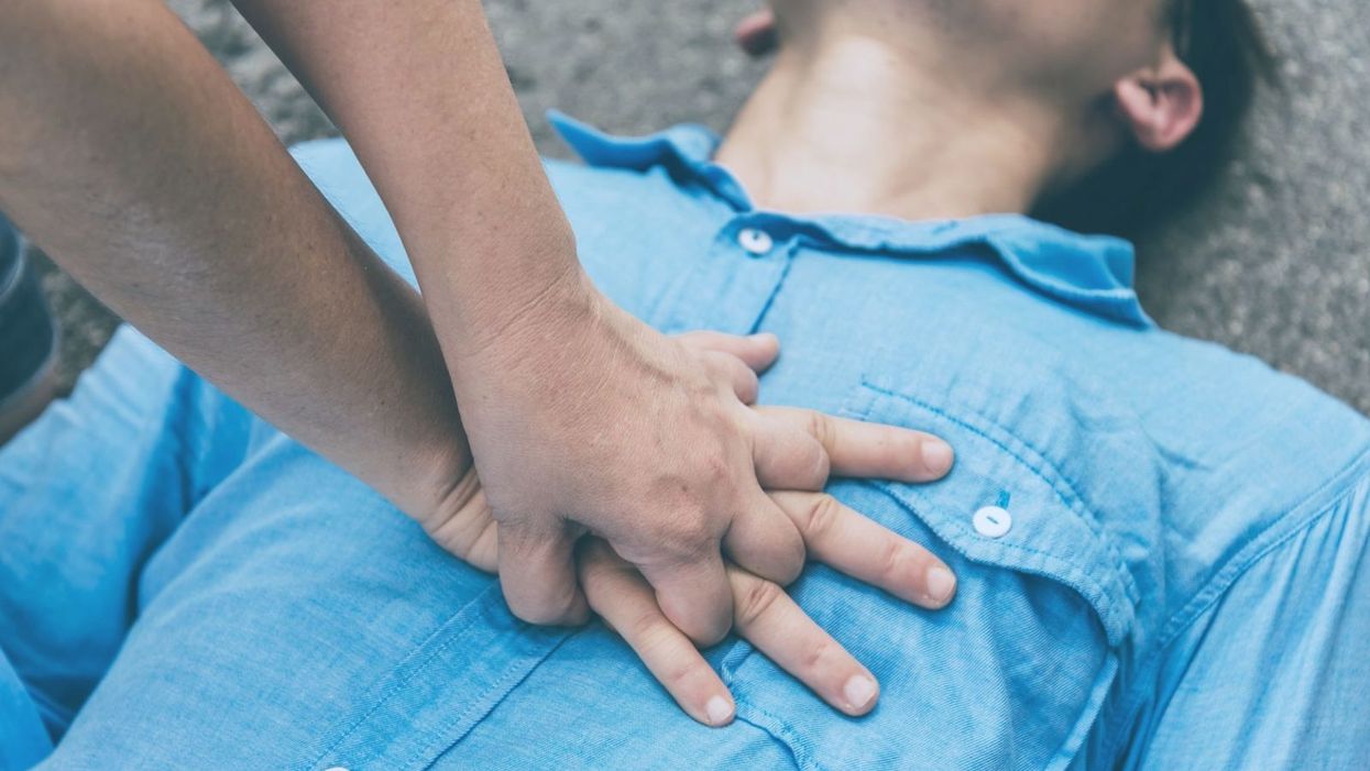
A man receiving cardiopulmonary resuscitation (CPR).
(Photo credit: spkphotostock/Adobe)
Last year, there were widespread reports of a 53-year-old Frenchman who had suffered a cardiac arrest and "died," but was then resuscitated back to life 18 hours after his heart had stopped.
The once black-and-white line between life and death is now blurrier than ever.
This was thought to have been possible in part because his body had progressively cooled down naturally after his heart had stopped, through exposure to the outside cold. The medical team who revived him were reported as being "stupefied" that they had been able to bring him back to life, in particular since he had not even suffered brain damage.
Interestingly, this man represents one of a growing number of extraordinary cases in which people who would otherwise be declared dead have now been revived. It is a testament to the incredible impact of resuscitation science -- a science that is providing opportunities to literally reverse death, and in doing so, shedding light on the age-old question of what happens when we die.
Death: Past and Present
Throughout history, the boundary between life and death was marked by the moment a person's heart stopped, breathing ceased, and brain function shut down. A person became motionless, lifeless, and was deemed irreversibly dead. This is because once the heart stops beating, blood flow stops and oxygen is cut off from all the body's organs, including the brain. Consequently, within seconds, breathing stops and brain activity comes to a halt. Since the cessation of the heart literally occurs in a "moment," the philosophical notion of a specific point in time of "irreversible" death still pervades society today. The law, for example, relies on "time of death," which corresponds to when the heart stops beating.
The advent of cardiopulmonary resuscitation (CPR) in the 1960s was revolutionary, demonstrating that the heart could potentially be restarted after it had stopped, and what had been a clear black-and-white line was shown to be potentially reversible in some people. What was once called death—the ultimate end point— was now widely called cardiac arrest, and became a starting point.
From then on, it was only if somebody had requested not to be resuscitated or when CPR was deemed to have failed that people would be declared dead by "cardiopulmonary criteria." Biologically, cardiac arrest and death by cardiopulmonary criteria are the same process, albeit marked at different points in time depending on when a declaration of death is made.
The apparent irreversibility of death as we know it may not necessarily reflect true irretrievable cellular damage inside the body.
Clearly, contrary to many people's perceptions, cardiac arrest is not a heart attack; it is the final step in death irrespective of cause, whether it be a stroke, a heart attack, a car accident, an overwhelming infection or cancer. This is how roughly 95 percent of the population are declared dead.
The only exception is the small proportion of people who may have suffered catastrophic brain injuries, but whose hearts can be artificially kept beating for a period of time on life-support machines. These people can be legally declared dead based on brain death criteria before their hearts have stopped. This is because the brain can die either from oxygen starvation after cardiac arrest or from massive trauma and internal bleeding. Either way, the brain dies hours or possibly longer after these injuries have taken place and not just minutes.
A Profound Realization
What has become increasingly clear is that the apparent irreversibility of death as we know it may not necessarily reflect true irretrievable cellular damage inside the body. This is consistent with a mounting understanding: it is only after a person actually dies that the cells in the body start to undergo their own process of death. Intriguingly, this process is something that can now be manipulated through medical intervention. Being cold is one of the factors that slows down the rate of cellular decay. The 53-year-old Frenchman's case and the other recent cases of resuscitation after prolonged periods of time illustrate this new understanding.
Last week's earth-shattering announcement by neuroscientist Dr. Nenad Sestan and his team out of Yale, published in the prestigious scientific journal Nature, provides further evidence that a time gap exists between actual death and cellular death in cadavers. In this seminal study, these researchers were able to restore partial function in pig brains four hours after their heads were severed from their bodies. These results follow from the pioneering work in 2001 of geneticist Fred Gage and colleagues from the Salk Institute, also published in Nature, which demonstrated the possibility of growing human brain cells in the laboratory by taking brain biopsies from cadavers in the mortuary up to 21 hours post-mortem.
The once black-and-white line between life and death is now blurrier than ever. Some people may argue this means these humans and pigs weren't truly "dead." However, that is like saying the people who were guillotined during the French Revolution were also not dead. Clearly, that is not the case. They were all dead. The problem is not death; it's our reliance on an outdated philosophical, rather than biological, notion of death.
Death can no longer be considered an absolute moment but rather a process that can be reversed even many hours after it has taken place.
But the distinction between irreversibility from a medical perspective and biological irreversibility may not matter much from a pragmatic perspective today. If medical interventions do not exist at any given time or place, then of course death cannot be reversed.
However, it is crucial to distinguish between biologically and medically: When "irreversible" loss of function arises due to inadequate treatment, then a person could be potentially brought back in the future when an alternative therapy becomes available, or even today if he or she dies in a location where novel treatments can slow down the rate of cell death. However, when true irreversible loss of function arises from a biological perspective, then no treatment will ever be able to reverse the process, whether today, tomorrow, or in a hundred years.
Probing the "Grey Zone"
Today, thanks to modern resuscitation science, death can no longer be considered an absolute moment but rather a process that can be reversed even many hours after it has taken place. How many hours? We don't really know.
One of the wider implications of our medical advances is that we can now study what happens to the human mind and consciousness after people enter the "grey zone," which marks the time after the heart stops, but before irreversible and irretrievable cell damage occurs, and people are then brought back to life. Millions have been successfully revived and many have reported experiencing a unique, universal, and transformative mental state.
Were they "dead"? Yes, according to all the criteria we have ever used. But they were able to be brought back before their "dead" bodies had reached the point of permanent, irreversible cellular damage. This reflects the period of death for all of us. So rather than a "near-death experience," I prefer a new terminology to describe these cases -- "an actual-death experience." These survivors' unique experiences are providing eyewitness testimonies of what we will all be likely to experience when we die.
Such an experience reportedly includes seeing a warm light, the presence of a compassionate perfect individual, deceased relatives, a review of their lives, a judgment of their actions and intentions as they pertain to their humanity, and in some cases a sensation of seeing doctors and nurses working to resuscitate them.
Are these experiences compatible with hallucinations or illusions? No -- in part, because these people have described real, verifiable events, which, by definition are not hallucinations, and in part, because their experiences are not compatible with confused and delirious memories that characterize oxygen deprivation.
The challenge for us scientifically is understanding how this is possible at a time when all our science tells us the brain shuts down.
For instance, it is hard to classify a structured meaningful review of one's life and one's humanity as hallucinatory or illusory. Instead, these experiences represent a new understanding of the overall human experience of death. As an intensive care unit physician for more than 10 years, I have seen numerous cases where these reports have been corroborated by my colleagues. In short, these survivors have been known to come back with reports of full consciousness, with lucid, well-structured thought processes and memory formation.
The challenge for us scientifically is understanding how this is possible at a time when all our science tells us the brain shuts down. The fact that these experiences occur is a paradox and suggests the undiscovered entity we call the "self," "consciousness," or "psyche" – the thing that makes us who we are - may not become annihilated at the point of so-called death.
At New York University, the State University of New York, and across 20 hospitals in the U.S. and Europe, we have brought together a new multi-disciplinary team of experts across many specialties, including neurology, cardiology, and intensive care. Together, we hope to improve cardiac arrest prevention and treatment, as well as to address the impact of new scientific discoveries on our understanding of what happens at death.
One of our first studies, Awareness during Resuscitation (AWARE), published in the medical journal Resuscitation in 2014, confirmed that some cardiac arrest patients report a perception of awareness without recall; others report detailed memories and experiences; and a few report full auditory and visual awareness and consciousness of their experience, from a time when brain function would be expected to have ceased.
While you probably have some opinion or belief about this based upon your own philosophical, religious, or cultural background, you may not realize that exploring what happens when we die is now a subject that science is beginning to investigate.
There is no question more intriguing to humankind. And for the first time in our history, we may finally uncover some real answers.
Dr. Sam Parnia MD, PhD is an Associate Professor of Critical Care Medicine at New York University School of Medicine, where he directs the Critical Care and Resuscitation Research Science Center. One of the world's leading experts on cardiac arrest resuscitation, post-cardiac arrest syndrome, and the scientific study of death, Dr. Parnia’s research focus is on developing new methods to save the lives and brains of patients who undergo cardiac arrest, as well as shedding light on what happens to our brains when we die. He also founded and directed the Human Consciousness Project, which featured an international consortium of scientists and physicians researching the nature of consciousness and its relationship to the brain during cardiac arrest. Dr. Parnia is also the author of two popular books, “What Happens When We Die?” and The New York Times bestseller, “Erasing Death: The Science that is Rewriting the Boundaries between Life and Death.”
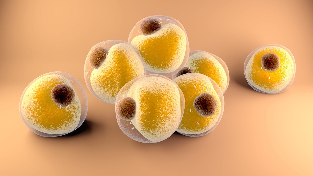
Researchers at Stanford have found that the virus that causes Covid-19 can infect fat cells, which could help explain why obesity is linked to worse outcomes for those who catch Covid-19.
Adobe Stock
Obesity is a risk factor for worse outcomes for a variety of medical conditions ranging from cancer to Covid-19. Most experts attribute it simply to underlying low-grade inflammation and added weight that make breathing more difficult.
Now researchers have found a more direct reason: SARS-CoV-2, the virus that causes Covid-19, can infect adipocytes, more commonly known as fat cells, and macrophages, immune cells that are part of the broader matrix of cells that support fat tissue. Stanford University researchers Catherine Blish and Tracey McLaughlin are senior authors of the study.
Most of us think of fat as the spare tire that can accumulate around the middle as we age, but fat also is present closer to most internal organs. McLaughlin's research has focused on epicardial fat, “which sits right on top of the heart with no physical barrier at all,” she says. So if that fat got infected and inflamed, it might directly affect the heart.” That could help explain cardiovascular problems associated with Covid-19 infections.
Looking at tissue taken from autopsy, there was evidence of SARS-CoV-2 virus inside the fat cells as well as surrounding inflammation. In fat cells and immune cells harvested from health humans, infection in the laboratory drove "an inflammatory response, particularly in the macrophages…They secreted proteins that are typically seen in a cytokine storm” where the immune response runs amok with potential life-threatening consequences. This suggests to McLaughlin “that there could be a regional and even a systemic inflammatory response following infection in fat.”
It is easy to see how the airborne SARS-CoV-2 virus infects the nose and lungs, but how does it get into fat tissue? That is a mystery and the source of ample speculation.
The macrophages studied by McLaughlin and Blish were spewing out inflammatory proteins, While the the virus within them was replicating, the new viral particles were not able to replicate within those cells. It was a different story in the fat cells. “When [the virus] gets into the fat cells, it not only replicates, it's a productive infection, which means the resulting viral particles can infect another cell,” including microphages, McLaughlin explains. It seems to be a symbiotic tango of the virus between the two cell types that keeps the cycle going.
It is easy to see how the airborne SARS-CoV-2 virus infects the nose and lungs, but how does it get into fat tissue? That is a mystery and the source of ample speculation.
Macrophages are mobile; they engulf and carry invading pathogens to lymphoid tissue in the lymph nodes, tonsils and elsewhere in the body to alert T cells of the immune system to the pathogen. Perhaps some of them also carry the virus through the bloodstream to more distant tissue.
ACE2 receptors are the means by which SARS-CoV-2 latches on to and enters most cells. They are not thought to be common on fat cells, so initially most researchers thought it unlikely they would become infected.
However, while some cell receptors always sit on the surface of the cell, other receptors are expressed on the surface only under certain conditions. Philipp Scherer, a professor of internal medicine and director of the Touchstone Diabetes Center at the University of Texas Southwestern Medical Center, suggests that, in people who have obesity, “There might be higher levels of dysfunctional [fat cells] that facilitate entry of the virus,” either through transiently expressed ACE2 or other receptors. Inflammatory proteins generated by macrophages might contribute to this process.
Another hypothesis is that viral RNA might be smuggled into fat cells as cargo in small bits of material called extracellular vesicles, or EVs, that can travel between cells. Other researchers have shown that when EVs express ACE2 receptors, they can act as decoys for SARS-CoV-2, where the virus binds to them rather than a cell. These scientists are working to create drugs that mimic this decoy effect as an approach to therapy.
Do fat cells play a role in Long Covid? “Fat cells are a great place to hide. You have all the energy you need and fat cells turn over very slowly; they have a half-life of ten years,” says Scherer. Observational studies suggest that acute Covid-19 can trigger the onset of diabetes especially in people who are overweight, and that patients taking medicines to regulate their diabetes “were actually quite protective” against acute Covid-19. Scherer has funding to study the risks and benefits of those drugs in animal models of Long Covid.
McLaughlin says there are two areas of potential concern with fat tissue and Long Covid. One is that this tissue might serve as a “big reservoir where the virus continues to replicate and is sent out” to other parts of the body. The second is that inflammation due to infected fat cells and macrophages can result in fibrosis or scar tissue forming around organs, inhibiting their function. Once scar tissue forms, the tissue damage becomes more difficult to repair.
Current Covid-19 treatments work by stopping the virus from entering cells through the ACE2 receptor, so they likely would have no effect on virus that uses a different mechanism. That means another approach will have to be developed to complement the treatments we already have. So the best advice McLaughlin can offer today is to keep current on vaccinations and boosters and lose weight to reduce the risk associated with obesity.
Keep Reading
Keep Reading
Bob Roehr is a biomedical journalist based in Washington, DC. Over the last twenty-five years he has written extensively for The BMJ, Scientific American, PNAS, Proto, and myriad other publications. He is primarily interested in HIV, infectious disease, immunology, and how growing knowledge of the microbiome is changing our understanding of health and disease. He is working on a book about the ways the body can at least partially control HIV and how that has influenced (or not) the search for a treatment and cure.
October 18 | 2022
Air pollution can lead to lung cancer. The connection suggests new ways to stop cancer in its tracks.
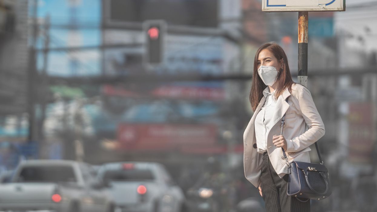
Researchers at Francis Crick Institute found that air pollution can "wake up" existing mutations, triggering them to turn into lung cancer. The scientists used their new understanding about this connection to prevent cancer in mice.
Adobe Stock
Forget taking a deep breath. Around the world, 99 percent of people breathe air polluted to unsafe levels, according to data from the World Health Organization. Activities such as burning fossil fuels release greenhouse gases that contribute to air pollution, which could lead to heart disease, stroke, asthma, emphysema, and some types of cancer.
“The burden of disease attributable to air pollution is now estimated to be on a par with other major global health risks such as unhealthy diet and tobacco smoking, and air pollution is now recognized as the single biggest environmental threat to human health,” wrote the authors of a 2021 WHO report.
The majority of lung cancer is attributed to smoking. But as pollution levels have increased, and anti-smoking campaigns have discouraged smoking, the proportion of lung cancers diagnosed in non-smokers has grown. The CDC estimates that 10 to 20 percent of lung cancers in the U.S. currently occur in non-smokers.
The mechanism between air pollution and the development of lung cancer has been unclear, but researchers at London’s Francis Crick Institute recently made an important breakthrough in understanding the connection. Lead investigator Charles Swanton presented this research last month at a conference in Paris.
Pollution awakens mutations
The Crick Institute scientists were able to identify a new link between common air pollutants and non-small cell lung cancer (NSCLC). They focused on pollutants called particulate matter, or PM, that are 2.5 microns wide, narrower than human cells.
Most cancer diagnosed in non-smoking people is NSCLC, but this type of cancer hasn’t received the same research attention as more common lung cancers found in smokers, according to Clare Weeden, a cancer researcher at the Crick Institute and a co-author of the study.
“This is a really underserved and under-researched population that we really need to tackle, as well as lung cancers that occur in smokers,” she says. “Lung cancer is the number one cancer killer worldwide.”
In the past, some researchers believed air pollution caused mutations that led to cancer. Others believed these mutations could remain dormant without any detriment to health until pollutants or other stressors triggered them to become cancerous. Reviving the latter hypothesis that carcinogens may activate pre-existing mutations, instead of directly causing them, the Crick researchers analyzed samples from 463,679 people in the UK and parts of Asia, noting mutations and comparing changes in gene expression in mice and human cells.
“The mutation can exist in a nascent clone without causing cancer,” says Emilia Lim, a bioinformatics expert and a co-first author of the Crick study. “It is the carcinogen that promotes a conducive environment for this one little clone to grow and expand into cancer. Through our work, we were able to revive excitement for this hypothesis and bring it to light.”
The study explains a confusing pattern of lung cancer developing, particularly in women, despite a lack of environmental risk factors like smoking, secondhand smoke, or radon exposure. The culprit in these cases may have been too much PM 2.5 exposure.
Other researchers had previously identified a link between mutations in certain genes that control epidermal growth factor receptors, or EGFR mutations, and the development of NSCLC. In a 2019 study of 250 people with this type of cancer, about 32 percent had the mutation. Women are more likely to have EGFR mutations than men.
Not everyone who has the EGFR mutation will develop lung cancer. Respirologist Stephen Lam studies lung cancer at the BC Cancer Research Centre in Vancouver, Canada, but was not involved in the Crick Institute research. He says the study explains a confusing pattern of lung cancer developing, particularly in women, despite a lack of environmental risk factors like smoking, secondhand smoke, or radon exposure. The culprit in these cases may have been too much PM 2.5 exposure.
More exposure leads to inflammation and lesions
The Crick researchers found that an excess of PM 2.5 in the air sparked an inflammatory process in cells within the lung. This inflammation set the stage for NSCLC to develop in people and mice with existing EGFR mutations.
The researchers also exposed mice without EGFR mutations to PM 2.5 pollution—an experiment that couldn’t be ethically conducted in humans—to link pollution exposure to NSCLC. The mice experiments also showed that NSCLC is dose-dependent; higher levels of exposure were associated with higher number of cancerous lesions forming.
Ultimately, the study “fundamentally changed how we view lung cancer in people who have never smoked,” said Swanton in a Crick Institute press release. “Cells with cancer-causing mutations accumulate naturally as we age, but they are normally inactive. We’ve demonstrated that air pollution wakes these cells up in the lungs, encouraging them to grow and potentially form tumors.”
Preventing cancer before it begins
Targeted therapies already exist for people with EGFR mutations who’ve developed NSCLC, but they have many side effects, according to Weeden. Researchers hope that making more definitive links between pollutants and cancer could help prevent people with EGFR or other mutations from developing lung cancer in the first place.
Along those lines, as an additional component of their study presented last month, the Crick researchers were able to prevent cancer in mice that had the EGFR mutations by blocking inflammation. They used an antibody to inhibit a protein called interleukin 1 beta, which plays a key role in inflammation. Scientists could eventually use such antibodies or other therapies to make a drug treatment that people can take to stop cancer in its tracks, even if they live in highly polluted areas.
Such potential could reach beyond lung cancer; in the past, Crick and other researchers have also found associations between exposure to air pollution and mesothelioma, as well as cancers of the small intestine, lip, mouth, and throat. These links could be meaningful to a growing number of people as climate change intensifies, and with increases in air pollution from fossil fuel combustion and natural disasters like forest fires.
Plus, air pollution is just one external condition that can flip the switch of these inflammatory pathways. Identifying a link between pollution and cancer “has wide ramifications for many other environmental factors that may [play] similar roles,” Weedon says. She hopes that the Crick study and future research in this area will offer some hope for non-smokers frustrated by cancer diagnoses.
Keep Reading
Keep Reading
Robin Donovan is a science journalist based in Portland, Oregon. Her work has appeared in Vice, Neo.Life, The Scientist, Willamette Week and many other outlets.