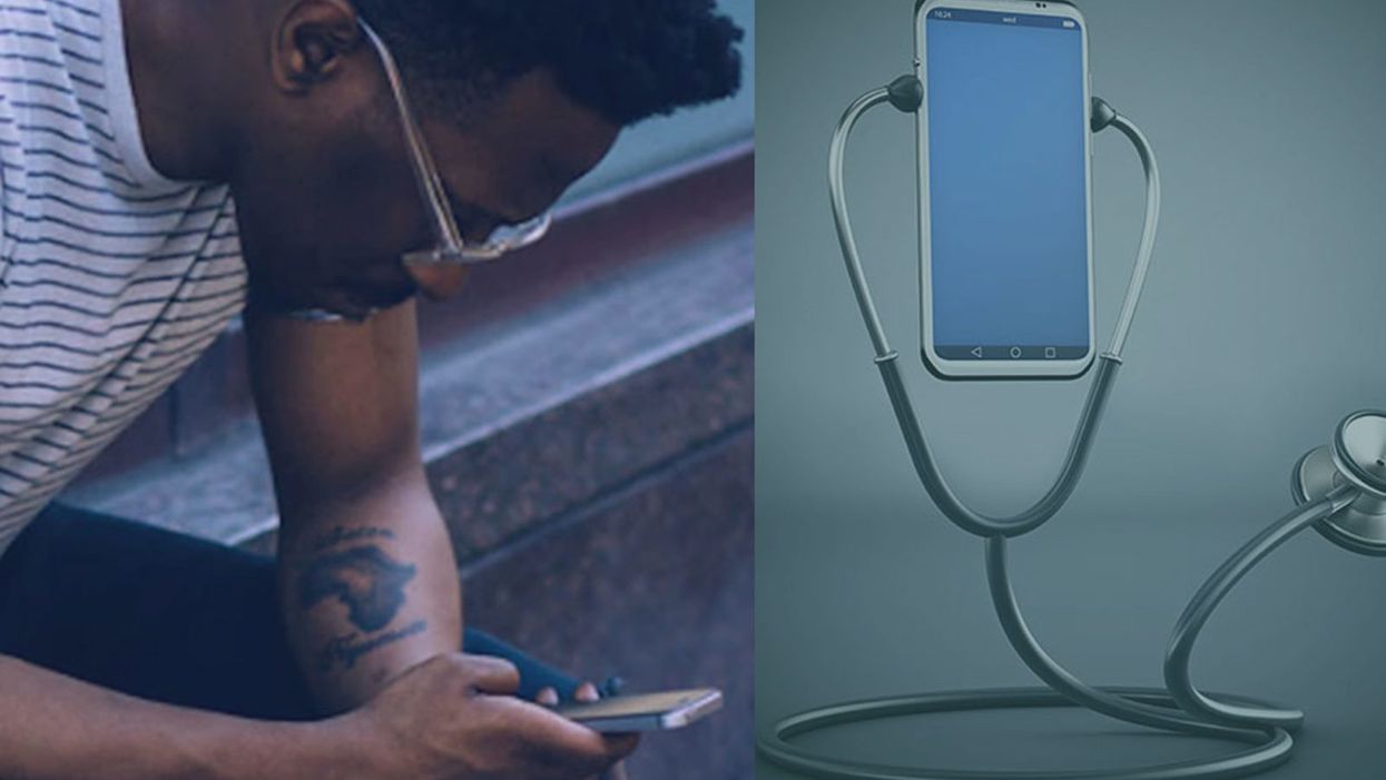
Diagnosed by App: Medical Testing in the Palm of Your Hand

The shift to in-home medical testing through smartphone apps is expected to save patients time in receiving certain kinds of diagnoses, but raises some privacy concerns.
Urinary tract infections aren't life-threatening, but they can be excruciatingly painful and debilitating.
"Overnight, I'd be gripped by this searing pain and I can barely walk," says Ling Koh, a Los Angeles-based bioengineer. But short of going to the ER or urgent care, she'd have to suffer for a few days until she could get in to see her family doctor for an antibiotic prescription.
Smartphones are now able to do on-the-spot diagnostic tests that were previously only able to be performed in a lab.
No longer. Koh, who works for Scanwell Health, was instrumental in the development of the company's smartphone app that is FDA-cleared for urinary tract infection screening. It allows someone to test urine at home using a paper test strip — the same one used by doctors in ERs and labs. The phone app reads a scan card from the test kit that can analyze what's on the strip and then connect the patient to a physician who can make a virtual diagnosis.
Test strips cost $15 for a three-pack and consultation with a doc is about the same as an average co-pay -- $25, and the app matches the quality of clinical laboratory tests, according to the company. Right now, you can get a referral to a telehealth visit with a doctor in California and get a prescription. A national rollout is in the works within the next couple of months.
"It's so easy to use them at home and eliminate the inefficiencies in the process," says Koh. "A telemedicine doctor can look at the test results and prescribe directly to the pharmacy instead of women waiting at home, miserable, and crying in the bathtub."
Scanwell is now involved in an ongoing National Institutes of Health- sponsored study of chronic kidney disease to test a version of the app to identify patients who have the disease, which affects more than 30 million Americans. "Because kidney disease has virtually no symptoms, by the time people realize they're sick, their illness is advanced and they're ready for dialysis," says Koh. "If we can catch it sooner, early intervention can help them avoid kidney failure."
Smartphones have changed society — and now they may change medical care, too. Thanks to the incredible processing capabilities of our smartphones, which come equipped with a camera, access to the internet and are thousands of times faster than the 1960s era NASA computers that ran the Apollo Moon Mission, these pocket-sized powerhouses have become an invaluable tool for managing our health and are even able to do on-the-spot diagnostic tests that were previously only able to be performed in a lab.
This shift to in-home testing is the wave of the future, promising to ease some of the medical care bottlenecks in which patients can have two- to three-week waits to see their family doctors and lift some of the burdens on overworked physicians.
"This is really the democratization of medicine because a lot of the things we used to rely on doctors, hospitals, or labs to do we'll be able to do ourselves," says Dr. Eric Topol, an eminent cardiologist and digital health pioneer at the Scripps Clinic and Research Institute in La Jolla.
But troubling questions remain. Aside from the obvious convenience, are these tests truly as accurate as ones in a doctor's office? And with all this medical information stored and collected by smartphones, will privacy be sacrificed? Will friends, family members, and employers suddenly have access to personal medical information we'd rather keep to ourselves?
The range of what these DIY health care apps can do is mind-boggling, and even more complex tests are on the way.
"I'm really worried about that because we've let our guard down," says Topol. "Data stored on servers is a target for cyber thieves — and data is being breached, hacked, brokered, and sold, and we're complacent."
Still, the apps have come a long way since 2011 when Topol whipped out an experimental smartphone electro-cardiogram that he had been testing on his patients when a fellow passenger on a flight from Washington D.C. was seized with severe chest pains. At 35,000 feet in the air, the app, which uses fingertip sensors to detect heart rate, showed the man was having a heart attack. After an emergency landing, the passenger was rushed to the closest hospital and survived. These days, even the Apple Watch has an FDA-approved app that can monitor your electro-cardiogram readings.
The range of what these DIY health care apps can do is mind-boggling, and even more complex tests are on the way. Phone apps can now monitor sleep quality to detect sleep apnea, blood pressure, weight and temperature. In the future, rapid diagnostic tests for infectious diseases, like flu, Dengue or Zika, and urinalysis will become common.
"There is virtually no limit to the kinds of testing that can be done using a smartphone," says Dr. John Halamka, Executive Director of the Health Technology Exploration Center at Beth Israel Lahey Health. "No one wants to drive to a clinician's office or lab if that same quality testing can be achieved at a lower cost without leaving home."
SkinVision's skin cancer screening tool, for instance, can tell if a suspicious mole is cancerous. Users take three photos, which are then run through the app's algorithm that compares their lesions with more than three million pictures, evaluating such elements as asymmetry, color, and shape, and spits out an assessment within thirty seconds. A team of in-house experts provide a review regardless of whether the mole is high or low risk, and the app encourages users to see their doctors. The Dutch-based company's app has been used by more than a million people globally in the EU, and in New Zealand and Australia, where skin cancer is rampant and early detection can save lives. The company has plans to enter the U.S. market, according to a spokesperson.
Apps like Instant Heart Rate analyze blood flow, which can indicate whether your heart is functioning normally, while uChek examines urine samples for up to 10 markers for conditions like diabetes and urinary tract infections. Some behavioral apps even have sensors that can spot suicide risks if users are less active, indicating they may be suffering from a bout of the blues.
Even more complex tests are in the research pipeline. Apps like ResAppDX could eventually replace x-rays, CT scans, and blood tests in diagnosing severe respiratory infections in kids, while an EU-funded project called i-Prognosis can track a variety of clues — voice changes, facial expressions, hand steadiness — that indicate the onset of Parkinson's disease.
These hand-held testing devices can be especially helpful in developing countries, and there are pilot programs to use smartphone technology to diagnose malaria and HIV infections in remote outposts in Africa.
"In a lot of these places, there's no infrastructure but everyone has a smartphone," says Scanwell's Koh. "We need to leverage the smartphone in a clinically relevant way."
However, patient privacy is an ongoing concern. A 2019 review in the Journal of the American Medical Association conducted by Australian and American researchers looked at three dozen behavioral health apps, mainly for depression and smoking cessation. They found that about 70 percent shared data with third parties, like Facebook and Google, but only one third of them disclosed this in a privacy policy.
"Patients just blindly accept the end user agreements without understanding the implications."
Users need to be vigilant, too. "Patients just blindly accept the end user agreements without understanding the implications," says Hamalka, who is also the Chief Information Officer and Dean for Technology at Harvard Medical School.
And quality control is an issue. Right now, the diagnostic tools currently available have been vetted by the FDA, and overseas companies like Skin Vision have been scrutinized by the U.K.'s National Health Service and the EU. But the danger is that a lot of apps are going to be popping up soon that haven't been properly tested, due to loopholes in the regulations.
"All we want," says Topol, "are rigorous studies to make sure what consumers are using is validated."
[Correction, August 19th, 2019: An earlier version of this story misstated the specifics of SkinVision's service. A team of in-house experts reviews users' submissions, not in-house dermatologists, and the service is not free.]
A new type of cancer therapy is shrinking deadly brain tumors with just one treatment
MRI scans after a new kind of immunotherapy for brain cancer show remarkable progress in one patient just days after the first treatment.
Few cancers are deadlier than glioblastomas—aggressive and lethal tumors that originate in the brain or spinal cord. Five years after diagnosis, less than five percent of glioblastoma patients are still alive—and more often, glioblastoma patients live just 14 months on average after receiving a diagnosis.
But an ongoing clinical trial at Mass General Cancer Center is giving new hope to glioblastoma patients and their families. The trial, called INCIPIENT, is meant to evaluate the effects of a special type of immune cell, called CAR-T cells, on patients with recurrent glioblastoma.
How CAR-T cell therapy works
CAR-T cell therapy is a type of cancer treatment called immunotherapy, where doctors modify a patient’s own immune system specifically to find and destroy cancer cells. In CAR-T cell therapy, doctors extract the patient’s T-cells, which are immune system cells that help fight off disease—particularly cancer. These T-cells are harvested from the patient and then genetically modified in a lab to produce proteins on their surface called chimeric antigen receptors (thus becoming CAR-T cells), which makes them able to bind to a specific protein on the patient’s cancer cells. Once modified, these CAR-T cells are grown in the lab for several weeks so that they can multiply into an army of millions. When enough cells have been grown, these super-charged T-cells are infused back into the patient where they can then seek out cancer cells, bind to them, and destroy them. CAR-T cell therapies have been approved by the US Food and Drug Administration (FDA) to treat certain types of lymphomas and leukemias, as well as multiple myeloma, but haven’t been approved to treat glioblastomas—yet.
CAR-T cell therapies don’t always work against solid tumors, such as glioblastomas. Because solid tumors contain different kinds of cancer cells, some cells can evade the immune system’s detection even after CAR-T cell therapy, according to a press release from Massachusetts General Hospital. For the INCIPIENT trial, researchers modified the CAR-T cells even further in hopes of making them more effective against solid tumors. These second-generation CAR-T cells (called CARv3-TEAM-E T cells) contain special antibodies that attack EFGR, a protein expressed in the majority of glioblastoma tumors. Unlike other CAR-T cell therapies, these particular CAR-T cells were designed to be directly injected into the patient’s brain.
The INCIPIENT trial results
The INCIPIENT trial involved three patients who were enrolled in the study between March and July 2023. All three patients—a 72-year-old man, a 74-year-old man, and a 57-year-old woman—were treated with chemo and radiation and enrolled in the trial with CAR-T cells after their glioblastoma tumors came back.
The results, which were published earlier this year in the New England Journal of Medicine (NEJM), were called “rapid” and “dramatic” by doctors involved in the trial. After just a single infusion of the CAR-T cells, each patient experienced a significant reduction in their tumor sizes. Just two days after receiving the infusion, the glioblastoma tumor of the 72-year-old man decreased by nearly twenty percent. Just two months later the tumor had shrunk by an astonishing 60 percent, and the change was maintained for more than six months. The most dramatic result was in the 57-year-old female patient, whose tumor shrank nearly completely after just one infusion of the CAR-T cells.
The results of the INCIPIENT trial were unexpected and astonishing—but unfortunately, they were also temporary. For all three patients, the tumors eventually began to grow back regardless of the CAR-T cell infusions. According to the press release from MGH, the medical team is now considering treating each patient with multiple infusions or prefacing each treatment with chemotherapy to prolong the response.
While there is still “more to do,” says co-author of the study neuro-oncologist Dr. Elizabeth Gerstner, the results are still promising. If nothing else, these second-generation CAR-T cell infusions may someday be able to give patients more time than traditional treatments would allow.
“These results are exciting but they are also just the beginning,” says Dr. Marcela Maus, a doctor and professor of medicine at Mass General who was involved in the clinical trial. “They tell us that we are on the right track in pursuing a therapy that has the potential to change the outlook for this intractable disease.”
A recent study in The Lancet Oncology showed that AI found 20 percent more cancers on mammogram screens than radiologists alone.
Since the early 2000s, AI systems have eliminated more than 1.7 million jobs, and that number will only increase as AI improves. Some research estimates that by 2025, AI will eliminate more than 85 million jobs.
But for all the talk about job security, AI is also proving to be a powerful tool in healthcare—specifically, cancer detection. One recently published study has shown that, remarkably, artificial intelligence was able to detect 20 percent more cancers in imaging scans than radiologists alone.
Published in The Lancet Oncology, the study analyzed the scans of 80,000 Swedish women with a moderate hereditary risk of breast cancer who had undergone a mammogram between April 2021 and July 2022. Half of these scans were read by AI and then a radiologist to double-check the findings. The second group of scans was read by two researchers without the help of AI. (Currently, the standard of care across Europe is to have two radiologists analyze a scan before diagnosing a patient with breast cancer.)
The study showed that the AI group detected cancer in 6 out of every 1,000 scans, while the radiologists detected cancer in 5 per 1,000 scans. In other words, AI found 20 percent more cancers than the highly-trained radiologists.

But even though the AI was better able to pinpoint cancer on an image, it doesn’t mean radiologists will soon be out of a job. Dr. Laura Heacock, a breast radiologist at NYU, said in an interview with CNN that radiologists do much more than simply screening mammograms, and that even well-trained technology can make errors. “These tools work best when paired with highly-trained radiologists who make the final call on your mammogram. Think of it as a tool like a stethoscope for a cardiologist.”
AI is still an emerging technology, but more and more doctors are using them to detect different cancers. For example, researchers at MIT have developed a program called MIRAI, which looks at patterns in patient mammograms across a series of scans and uses an algorithm to model a patient's risk of developing breast cancer over time. The program was "trained" with more than 200,000 breast imaging scans from Massachusetts General Hospital and has been tested on over 100,000 women in different hospitals across the world. According to MIT, MIRAI "has been shown to be more accurate in predicting the risk for developing breast cancer in the short term (over a 3-year period) compared to traditional tools." It has also been able to detect breast cancer up to five years before a patient receives a diagnosis.
The challenges for cancer-detecting AI tools now is not just accuracy. AI tools are also being challenged to perform consistently well across different ages, races, and breast density profiles, particularly given the increased risks that different women face. For example, Black women are 42 percent more likely than white women to die from breast cancer, despite having nearly the same rates of breast cancer as white women. Recently, an FDA-approved AI device for screening breast cancer has come under fire for wrongly detecting cancer in Black patients significantly more often than white patients.
As AI technology improves, radiologists will be able to accurately scan a more diverse set of patients at a larger volume than ever before, potentially saving more lives than ever.

