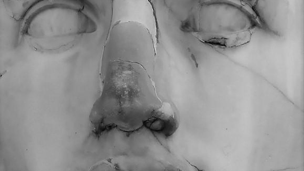
A Tool for Disease Detection Is Right Under Our Noses

In March, researchers published a review that lists which organic chemicals match up with certain diseases and biomarkers in the skin, saliva and urine. It’s an important step in creating a robot nose that can reliably detect diseases.
The doctor will sniff you now? Well, not on his or her own, but with a device that functions like a superhuman nose. You’ll exhale into a breathalyzer, or a sensor will collect “scent data” from a quick pass over your urine or blood sample. Then, AI software combs through an olfactory database to find patterns in the volatile organic compounds (VOCs) you secreted that match those associated with thousands of VOC disease biomarkers that have been identified and cataloged.
No further biopsy, imaging test or procedures necessary for the diagnosis. According to some scientists, this is how diseases will be detected in the coming years.
All diseases alter the organic compounds found in the body and their odors. Volatolomics is an emerging branch of chemistry that uses the smell of gases emitted by breath, urine, blood, stool, tears or sweat to diagnose disease. When someone is sick, the normal biochemical process is disrupted, and this alters the makeup of the gas, including a change in odor.
“These metabolites show a snapshot of what’s going on with the body,” says Cristina Davis, a biomedical engineer and associate vice chancellor of Interdisciplinary Research and Strategic Initiatives at the University of California, Davis. This opens the door to diagnosing conditions even before symptoms are present. It’s possible to detect a sweet, fruity smell in the breath of someone with diabetes, for example.
Hippocrates may have been the first to note that people with certain diseases give off an odor but dogs provided the proof of concept. Scientists have published countless studies in which dogs or other high-performing smellers like rodents have identified people with cancer, lung disease or other conditions by smell alone. The brain region that analyzes smells is proportionally about 40 times greater in dogs than in people. The noses of rodents are even more powerful.
Take prostate cancer, which is notoriously difficult to detect accurately with standard medical testing. After sniffing a tiny urine sample, trained dogs were able to pick out prostate cancer in study subjects more than 96 percent of the time, and earlier than a physician could in some cases.
But using dogs as bio-detectors is not practical. It is labor-intensive, complicated and expensive to train dogs to bark or lie down when they smell a certain VOC, explains Bruce Kimball, a chemical ecologist at the Monell Chemical Senses Center in Philadelphia. Kimball has trained ferrets to scratch a box when they smell a specific VOC so he knows. The lab animal must be taught to distinguish the VOC from background odors and trained anew for each disease scent.

In the lab of chemical ecologist Bruce Kimball, ferrets were trained to scratch a box when they identified avian flu in mallard ducks.
Glen J. Golden
There are some human super-smellers among us. In 2019, Joy Milne of Scotland proved she could unerringly identify people with Parkinson’s disease from a musky scent emitted from their skin. Clinical testing showed that she could distinguish the odor of Parkinson’s on a worn t-shirt before clinical symptoms even appeared.
Hossam Haick, a professor at Technion-Israel Institute of Technology, maintains that volatolomics is the future of medicine. Misdiagnosis and late detection are huge problems in health care, he says. “A precise and early diagnosis is the starting point of all clinical activities.” Further, this science has the potential to eliminate costly invasive testing or imaging studies and improve outcomes through earlier treatment.
The Nose Knows a Lot
“Volatolomics is not a fringe theory. There is science behind it,” Davis stresses. Every VOC has its own fingerprint, and a method called gas chromatography-mass spectrometry (GCMS) uses highly sensitive instruments to separate the molecules of these VOCs to determine their structures. But GCMS can’t discern the telltale patterns of particular diseases, and other technologies to analyze biomarkers have been limited.
We have technology that can see, hear and sense touch but scientists don’t have a handle yet on how smell works. The ability goes beyond picking out a single scent in someone’s breath or blood sample. It’s the totality of the smell—not the smell of a single chemical— which defines a disease. The dog’s brain is able to infer something when they smell a VOC that eludes human analysis so far.
Odor is a complex ecosystem and analyzing a VOC is compounded by other scents in the environment, says Kimball. A person’s diet and use of tobacco or alcohol also will affect the breath. Even fluctuations in humidity and temperature can contaminate a sample.
If successful, a sophisticated AI network can imitate how the dog brain recognizes patterns in smells. Early versions of robot noses have already been developed.
With today’s advances in data mining, AI and machine learning, scientists are trying to create mechanical devices that can draw on algorithms based on GCMS readings and data about diseases that dogs have sniffed out. If successful, a sophisticated AI network can imitate how the dog brain recognizes patterns in smells.
In March, Nano Research published a comprehensive review of volatolomics in health care authored by Haick and seven colleagues. The intent was to bridge gaps in the field for scientists trying to connect the biomarkers and sensor technology needed to develop a robot nose. This paper serves as a reference manual for the field that lists which VOCs are associated with what disease and the biomarkers in skin, saliva, breath, and urine.
Weiwei Wu, one of the co-authors and a professor at Xidian University in China, explains that creating a robotic nose requires the expertise of chemists, computer scientists, electrical engineers, material scientists, and clinicians. These researchers use different terms and methodologies and most have not collaborated before with the other disciplines. “The electrical engineers know the device but they don’t know as much about the biomarkers they need to detect,” Wu offers as an example.
This review is significant, Wu continues, because it can facilitate progress in the field by providing experts in all the disciplines with the basic knowledge needed to create an effective robot nose for diagnostic use. The paper also includes a systematic summary of the research methodology of volatolomics.
Once scientists build a stronger database of VOCs, they can program a device to identify critical patterns of specified diseases on a reliable basis. On a machine learning model, the algorithms automatically get better at diagnosing with each use. Wu envisions further tweaks in the next few years to make the devices smaller and consume less power.
A Whiff of the Future
Early versions of robot noses have already been developed. Some of them use chemical sensors to pick up smells in the breath or other body emission molecules. That data is sent through an electrical signal to a computer network for interpretation and possible linkage to a disease.
This electronic nose, or e-nose, has been successful in small pilot studies at labs around the world. At Ben-Gurion University in Israel, researchers detected breast cancer with electronic gas sensors with 95% accuracy, a higher sensitivity than mammograms. Other robot noses, called p-noses, use photons instead of electrical signals.
The mechanical noses being developed tap different methodologies and analytic techniques which makes it hard to compare them. Plus, the devices are intended for varying uses. One team, for example, is working on an e-nose that can be waved over a plate to screen for the presence of a particular allergen when you’re dining out.
A robot nose could be used as a real-time diagnostic tool in clinical practice. Kimball is working on one such tool that can distinguish between a viral and bacterial infection. This would enable physicians to determine whether an antibiotic prescription is appropriate without waiting for a lab result.
Davis is refining a hand-held device that identifies COVID-19 through a simple breath test. She sees the tool being used at crowded airports, sports stadiums and concert venues where PCR or rapid antigen testing is impractical. Background air samples are collected from the space so that those signals can be removed from the human breath measurement. “[The sensor tool] has the same accuracy as the rapid antigen test kits but exhaled breath is easier to collect,” she notes.

The NaNose, also known as the SniffPhone, uses tiny sensors boosted by AI to distinguish Alzheimer's, Crohn's disease, the early stages of several cancers, and other diseases with 84 to 98 percent accuracy.
Hossam Haick
Haick named his team’s robot nose, “NaNose,” since it is based on nanotechnology; the prototype is called the SniffPhone. Using tiny sensors boosted by AI, it can distinguish 23 diseases in human subjects with 84 to 98 percent accuracy. This includes early stages of several cancers, Alzheimer’s, tuberculosis and Crohn’s disease. His team has been raising the accuracy level by combining biomarker signals from both breath and skin, for example. The goal is to achieve 99.9 percent accuracy consistently so no other diagnostic tests would be needed before treating the patient. Plus, it will be affordable, he says.
Kimball predicts we’ll be seeing these diagnostic tools in the next decade. “The physician would narrow down what [the diagnosis] might be and then get the correct tool,” he says. Others are envisioning one device that can screen for multiple diseases by programming the software, which would be updated regularly with new findings.
Larger volatolomics studies must be conducted before these e-noses are ready for clinical use, however. Experts also need to learn how to establish normal reference ranges for e-nose readings to support clinicians using the tool.
“Taking successful prototypes from the lab to industry is the challenge,” says Haick, ticking off issues like reproducibility, mass production and regulation. But volatolomics researchers are unanimous in believing the future of health care is so close they can smell it.
A new type of cancer therapy is shrinking deadly brain tumors with just one treatment
MRI scans after a new kind of immunotherapy for brain cancer show remarkable progress in one patient just days after the first treatment.
Few cancers are deadlier than glioblastomas—aggressive and lethal tumors that originate in the brain or spinal cord. Five years after diagnosis, less than five percent of glioblastoma patients are still alive—and more often, glioblastoma patients live just 14 months on average after receiving a diagnosis.
But an ongoing clinical trial at Mass General Cancer Center is giving new hope to glioblastoma patients and their families. The trial, called INCIPIENT, is meant to evaluate the effects of a special type of immune cell, called CAR-T cells, on patients with recurrent glioblastoma.
How CAR-T cell therapy works
CAR-T cell therapy is a type of cancer treatment called immunotherapy, where doctors modify a patient’s own immune system specifically to find and destroy cancer cells. In CAR-T cell therapy, doctors extract the patient’s T-cells, which are immune system cells that help fight off disease—particularly cancer. These T-cells are harvested from the patient and then genetically modified in a lab to produce proteins on their surface called chimeric antigen receptors (thus becoming CAR-T cells), which makes them able to bind to a specific protein on the patient’s cancer cells. Once modified, these CAR-T cells are grown in the lab for several weeks so that they can multiply into an army of millions. When enough cells have been grown, these super-charged T-cells are infused back into the patient where they can then seek out cancer cells, bind to them, and destroy them. CAR-T cell therapies have been approved by the US Food and Drug Administration (FDA) to treat certain types of lymphomas and leukemias, as well as multiple myeloma, but haven’t been approved to treat glioblastomas—yet.
CAR-T cell therapies don’t always work against solid tumors, such as glioblastomas. Because solid tumors contain different kinds of cancer cells, some cells can evade the immune system’s detection even after CAR-T cell therapy, according to a press release from Massachusetts General Hospital. For the INCIPIENT trial, researchers modified the CAR-T cells even further in hopes of making them more effective against solid tumors. These second-generation CAR-T cells (called CARv3-TEAM-E T cells) contain special antibodies that attack EFGR, a protein expressed in the majority of glioblastoma tumors. Unlike other CAR-T cell therapies, these particular CAR-T cells were designed to be directly injected into the patient’s brain.
The INCIPIENT trial results
The INCIPIENT trial involved three patients who were enrolled in the study between March and July 2023. All three patients—a 72-year-old man, a 74-year-old man, and a 57-year-old woman—were treated with chemo and radiation and enrolled in the trial with CAR-T cells after their glioblastoma tumors came back.
The results, which were published earlier this year in the New England Journal of Medicine (NEJM), were called “rapid” and “dramatic” by doctors involved in the trial. After just a single infusion of the CAR-T cells, each patient experienced a significant reduction in their tumor sizes. Just two days after receiving the infusion, the glioblastoma tumor of the 72-year-old man decreased by nearly twenty percent. Just two months later the tumor had shrunk by an astonishing 60 percent, and the change was maintained for more than six months. The most dramatic result was in the 57-year-old female patient, whose tumor shrank nearly completely after just one infusion of the CAR-T cells.
The results of the INCIPIENT trial were unexpected and astonishing—but unfortunately, they were also temporary. For all three patients, the tumors eventually began to grow back regardless of the CAR-T cell infusions. According to the press release from MGH, the medical team is now considering treating each patient with multiple infusions or prefacing each treatment with chemotherapy to prolong the response.
While there is still “more to do,” says co-author of the study neuro-oncologist Dr. Elizabeth Gerstner, the results are still promising. If nothing else, these second-generation CAR-T cell infusions may someday be able to give patients more time than traditional treatments would allow.
“These results are exciting but they are also just the beginning,” says Dr. Marcela Maus, a doctor and professor of medicine at Mass General who was involved in the clinical trial. “They tell us that we are on the right track in pursuing a therapy that has the potential to change the outlook for this intractable disease.”
A recent study in The Lancet Oncology showed that AI found 20 percent more cancers on mammogram screens than radiologists alone.
Since the early 2000s, AI systems have eliminated more than 1.7 million jobs, and that number will only increase as AI improves. Some research estimates that by 2025, AI will eliminate more than 85 million jobs.
But for all the talk about job security, AI is also proving to be a powerful tool in healthcare—specifically, cancer detection. One recently published study has shown that, remarkably, artificial intelligence was able to detect 20 percent more cancers in imaging scans than radiologists alone.
Published in The Lancet Oncology, the study analyzed the scans of 80,000 Swedish women with a moderate hereditary risk of breast cancer who had undergone a mammogram between April 2021 and July 2022. Half of these scans were read by AI and then a radiologist to double-check the findings. The second group of scans was read by two researchers without the help of AI. (Currently, the standard of care across Europe is to have two radiologists analyze a scan before diagnosing a patient with breast cancer.)
The study showed that the AI group detected cancer in 6 out of every 1,000 scans, while the radiologists detected cancer in 5 per 1,000 scans. In other words, AI found 20 percent more cancers than the highly-trained radiologists.

But even though the AI was better able to pinpoint cancer on an image, it doesn’t mean radiologists will soon be out of a job. Dr. Laura Heacock, a breast radiologist at NYU, said in an interview with CNN that radiologists do much more than simply screening mammograms, and that even well-trained technology can make errors. “These tools work best when paired with highly-trained radiologists who make the final call on your mammogram. Think of it as a tool like a stethoscope for a cardiologist.”
AI is still an emerging technology, but more and more doctors are using them to detect different cancers. For example, researchers at MIT have developed a program called MIRAI, which looks at patterns in patient mammograms across a series of scans and uses an algorithm to model a patient's risk of developing breast cancer over time. The program was "trained" with more than 200,000 breast imaging scans from Massachusetts General Hospital and has been tested on over 100,000 women in different hospitals across the world. According to MIT, MIRAI "has been shown to be more accurate in predicting the risk for developing breast cancer in the short term (over a 3-year period) compared to traditional tools." It has also been able to detect breast cancer up to five years before a patient receives a diagnosis.
The challenges for cancer-detecting AI tools now is not just accuracy. AI tools are also being challenged to perform consistently well across different ages, races, and breast density profiles, particularly given the increased risks that different women face. For example, Black women are 42 percent more likely than white women to die from breast cancer, despite having nearly the same rates of breast cancer as white women. Recently, an FDA-approved AI device for screening breast cancer has come under fire for wrongly detecting cancer in Black patients significantly more often than white patients.
As AI technology improves, radiologists will be able to accurately scan a more diverse set of patients at a larger volume than ever before, potentially saving more lives than ever.

