
Often called the window to the soul, the eyes are more sacred than other body parts, at least for some.
Eye transplants are desperately needed, but they're nowhere in sight. About 12.7 million people worldwide need a corneal transplant, which means that only one in 70 people who require them, get them. The gaps are international. Eye banks in the United Kingdom are around 20 percent below the level needed to supply hospitals, while Indian eye banks, which need at least 250,000 corneas per year, collect only around 45 to 50 thousand donor corneas (and of those 60 to 70 percent are successfully transplanted).
As for retinas, it's impossible currently to put one into the eye of another person. Artificial devices can be implanted to restore the sight of patients suffering from severe retinal diseases, but the number of people around the world with such “bionic eyes” is less than 600, while in America alone 11 million people have some type of retinal disease leading to severe vision loss. Add to this an increasingly aging population, commonly facing various vision impairments, and you have a recipe for heavy burdens on individuals, the economy and society. In the U.S. alone, the total annual economic impact of vision problems was $51.4 billion in 2017.
Even if you try growing tissues in the petri dish route into organoids mimicking the function of the human eye, you will not get the physiological complexity of the structure and metabolism of the real thing, according to Cosma. She is a member of a scientific consortium that includes researchers from major institutions from Spain, the U.K., Portugal, Italy and Israel. The consortium has received about $3.8 million from the European Union to pursue innovative eye research. Her team’s goal is to give hope to at least 2.2 billion people across the world afflicted with a vision impairment and 33 million who go through life with avoidable blindness.
Their method? Resuscitating cadaveric eyes for at least a month.
If we succeed, it will be the first intact human model of the eye capable of exploring and analyzing regenerative processes ex vivo. -- Maria Pia Cosma.
“We proposed to resuscitate eyes, that is to restore the global physiology and function of human explanted tissues,” Cosma said, referring to living tissues extracted from the eye and placed in a medium for culture. Their ECaBox is an ex vivo biological system, in which eyes taken from dead donors are placed in an artificial environment, designed to preserve the eye’s temperature and pH levels, deter blood clots, and remove the metabolic waste and toxins that would otherwise spell their demise.
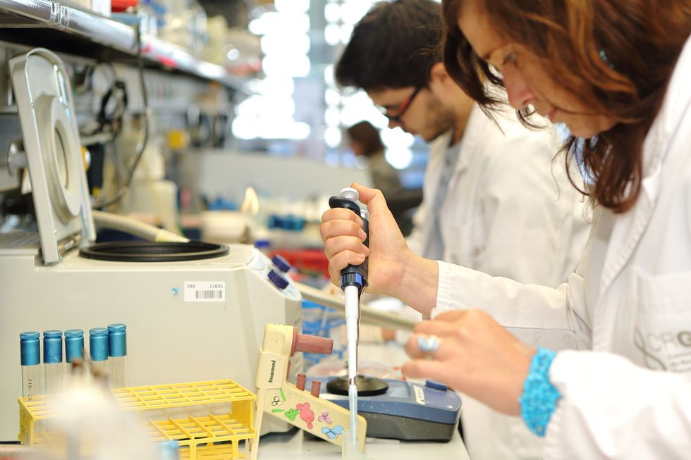
Scientists work on resuscitating eyes in the lab of Maria Pia Cosma.
Courtesy of Maria Pia Cosma.
“One of the great challenges is the passage of the blood in the capillary branches of the eye, what we call long-term perfusion,” Cosma said. Capillaries are an intricate network of very thin blood vessels that transport blood, nutrients and oxygen to cells in the body’s organs and systems. To maintain the garland-shaped structure of this network, sufficient amounts of oxygen and nutrients must be provided through the eye circulation and microcirculation. “Our ambition is to combine perfusion of the vessels with artificial blood," along with using a synthetic form of vitreous, or the gel-like fluid that lets in light and supports the the eye's round shape, Cosma said.
The scientists use this novel setup with the eye submersed in its medium to keep the organ viable, so they can test retinal function. “If we succeed, we will ensure full functionality of a human organ ex vivo. It will be the first intact human model of the eye capable of exploring and analyzing regenerative processes ex vivo,” Cosma added.
A rapidly developing field of regenerative medicine aims to stimulate the body's natural healing processes and restore or replace damaged tissues and organs. But for people with retinal diseases, regenerative medicine progress has been painfully slow. “Experiments on rodents show progress, but the risks for humans are unacceptable,” Cosma said.
The ECaBox could boost progress with regenerative medicine for people with retinal diseases, which has been painfully slow because human experiments involving their eyes are too risky. “We will test emerging treatments while reducing animal research, and greatly accelerate the discovery and preclinical research phase of new possible treatments for vision loss at significantly reduced costs,” Cosma explained. Much less time and money would be wasted during the drug discovery process. Their work may even make it possible to transplant the entire eyeball for those who need it.
“It is a very exciting project,” said Sanjay Sharma, a professor of ophthalmology and epidemiology at Queen's University, in Kingston, Canada. “The ability to explore and monitor regenerative interventions will increasingly be of importance as we develop therapies that can regenerate ocular tissues, including the retina.”
Seemingly, there's no sacred religious text or a holy book prohibiting the practice of eye donation.
But is the world ready for eye transplants? “People are a bit weird or very emotional about donating their eyes as compared to other organs,” Cosma said. And much can be said about the problem of eye donor shortage. Concerns include disfigurement and healthcare professionals’ fear that the conversation about eye donation will upset the departed person’s relatives because of cultural or religious considerations. As just one example, Sharma noted the paucity of eye donations in his home country, Canada.
Yet, experts like Sharma stress the importance of these donations for both the recipients and their family members. “It allows them some psychological benefit in a very difficult time,” he said. So why are global eye banks suffering? Is it because the eyes are the windows to the soul?
Seemingly, there's no sacred religious text or a holy book prohibiting the practice of eye donation. In fact, most major religions of the world permit and support organ transplantation and donation, and by extension eye donation, because they unequivocally see it as an “act of neighborly love and charity.” In Hinduism, the concept of eye donation aligns with the Hindu principle of daan or selfless giving, where individuals donate their organs or body after death to benefit others and contribute to society. In Islam, eye donation is a form of sadaqah jariyah, a perpetual charity, as it can continue to benefit others even after the donor's death.
Meanwhile, Buddhist masters teach that donating an organ gives another person the chance to live longer and practice dharma, the universal law and order, more meaningfully; they also dismiss misunderstandings of the type “if you donate an eye, you’ll be born without an eye in the next birth.” And Christian teachings emphasize the values of love, compassion, and selflessness, all compatible with organ donation, eye donation notwithstanding; besides, those that will have a house in heaven, will get a whole new body without imperfections and limitations.
The explanation for people’s resistance may lie in what Deepak Sarma, a professor of Indian religions and philosophy at Case Western Reserve University in Cleveland, calls “street interpretation” of religious or spiritual dogmas. Consider the mechanism of karma, which is about the causal relation between previous and current actions. “Maybe some Hindus believe there is karma in the eyes and, if the eye gets transplanted into another person, they will have to have that karmic card from now on,” Sarma said. “Even if there is peculiar karma due to an untimely death–which might be interpreted by some as bad karma–then you have the karma of the recipient, which is tremendously good karma, because they have access to these body parts, a tremendous gift,” Sarma said. The overall accumulation is that of good karma: “It’s a beautiful kind of balance,” Sarma said.
For the Jews, Christians, and Muslims who believe in the physical resurrection of the body that will be made new in an afterlife, the already existing body is sacred since it will be the basis of a new refashioned body in an afterlife.---Omar Sultan Haque.
With that said, Sarma believes it is a fallacy to personify or anthropomorphize the eye, which doesn’t have a soul, and stresses that the karma attaches itself to the soul and not the body parts. But for scholars like Omar Sultan Haque—a psychiatrist and social scientist at Harvard Medical School, investigating questions across global health, anthropology, social psychology, and bioethics—the hierarchy of sacredness of body parts is entrenched in human psychology. You cannot equate the pinky toe with the face, he explained.
“The eyes are the window to the soul,” Haque said. “People have a hierarchy of body parts that are considered more sacred or essential to the self or soul, such as the eyes, face, and brain.” In his view, the techno-utopian transhumanist communities (especially those in Silicon Valley) have reduced the totality of a person to a mere material object, a “wet robot” that knows no sacredness or hierarchy of human body parts. “But for the Jews, Christians, and Muslims who believe in the physical resurrection of the body that will be made new in an afterlife, the [already existing] body is sacred since it will be the basis of a new refashioned body in an afterlife,” Haque said. “You cannot treat the body like any old material artifact, or old chair or ragged cloth, just because materialistic, secular ideologies want so,” he continued.
For Cosma and her peers, however, the very definition of what is alive or not is a bit semantic. “As soon as we die, the electrophysiological activity in the eye stops,” she said. “The goal of the project is to restore this activity as soon as possible before the highly complex tissue of the eye starts degrading.” Cosma’s group doesn’t yet know when they will be able to keep the eyes alive and well in the ECaBox, but the consensus is that the sooner the better. Hopefully, the taboos and fears around the eye donations will dissipate around the same time.
Doctors used new eye drops to treat a rare genetic disorder.
The gene therapy: In May 2023, the FDA approved Vyjuvek, the first gene therapy to treat DEB.
Vyjuvek uses an inactivated herpes simplex virus to deliver working copies of the gene for collagen 7 to the body’s cells. In small trials, 65 percent of DEB-caused wounds sprinkled with it healed completely, compared to just 26 percent of wounds treated with a placebo.
“It was like looking through thick fog.” -- Antonio Vento Carvajal.
The patient: Antonio Vento Carvajal, a 14 year old living in Florida, was one of the trial participants to benefit from Vyjuvek, which was developed by Pittsburgh-based pharmaceutical company Krystal Biotech.
While the topical gene therapy could help his skin, though, it couldn’t do anything to address the severe vision loss Antonio experienced due to his DEB. He’d undergone multiple surgeries to have scar tissue removed from his eyes, but due to his condition, the blisters keep coming back.
“It was like looking through thick fog,” said Antonio, noting how his impaired vision made it hard for him to play his favorite video games. “I had to stand up from my chair, walk over, and get closer to the screen to be able to see.”
The idea: Encouraged by how Antonio’s skin wounds were responding to the gene therapy, Alfonso Sabater, his doctor at the Bascom Palmer Eye Institute, reached out to Krystal Biotech to see if they thought an alternative formula could potentially help treat his patient’s eyes.
The company was eager to help, according to Sabater, and after about two years of safety and efficacy testing, he had permission, under the FDA’s compassionate use protocol, to treat Antonio’s eyes with a version of the topical gene therapy delivered as eye drops.
The results: In August 2022, Sabater once again removed scar tissue from Antonio’s right eye, but this time, he followed up the surgery by immediately applying eye drops containing the gene therapy.
“I would send this message to other families in similar situations, whether it’s DEB or another condition that can benefit from genetic therapy. Don’t be afraid.” -- Yunielkys “Yuni” Carvajal.
The vision in Antonio’s eye steadily improved. By about eight months after the treatment, it was just slightly below average (20/25) and stayed that way. In March 2023, Sabater performed the same procedure on his young patient’s other eye, and the vision in it has also steadily improved.
“I’ve seen the transformation in Antonio’s life,” said Sabater. “He’s always been a happy kid. Now he’s very happy. He can function pretty much normally. He can read, he can study, he can play video games.”
Looking ahead: The topical gene therapy isn’t a permanent fix — it doesn’t alter Antonio’s own genes, so he has to have the eye drops reapplied every month. Still, that’s far less invasive than having to undergo repeated surgeries.
Sabater is now working with Krystal Biotech to launch trials of the eye drops in other patients, and not just those with DEB. By changing the gene delivered by the therapy, he believes it could be used to treat other eye disorders that are far more common — Fuchs’ dystrophy, for example, affects the vision of an estimated 300 million people over the age of 30.
Antonio’s mother, Yunielkys “Yuni” Carvajal, meanwhile, has said that having her son be the first to receive the eye drops was “very scary,” but she’s hopeful others will take a chance on new gene therapies if given the opportunity.
“I would send this message to other families in similar situations, whether it’s DEB or another condition that can benefit from genetic therapy,” she said. “Don’t be afraid.”
This article originally appeared on Freethink, home of the brightest minds and biggest ideas of all time.

A chatbot from the nonprofit National Eating Disorders Association aimed to provide 24/7 guidance on eating disorders. Several drawbacks point to the importance of therapist-tech collaboration in multiple areas of health.
Then everything went off the rails. Tessa told Maxwell an unequivocal yes, and that “it’s important to approach weight loss with a balanced and realistic mindset,” and recommended eating whole foods and lean proteins to create a 500-1000 calorie per day deficit that would lead to a loss of 1-2 pounds per week. To most people, the advice sounds anodyne, but alarm bells sounded in Maxwell’s head.
“This is actively going to feed eating disorders,” Maxwell says. “Having a chatbot be the direct response to someone reaching out for support for an eating disorder instead of the helpline seems careless.”
“The scripts that are being fed into the chatbot are only going to be as good as the person who’s feeding them.” -- Alexis Conason.
According to several decades of research, deliberate weight loss in the form of dieting is a serious risk for people with eating disorders. Maxwell says that following medical advice like what Tessa prescribed was what triggered her eating disorder as a child. And Maxwell wasn’t the only one who got such advice from the bot. When eating disorder therapist Alexis Conason tried Tessa, she asked the AI chatbot many of the questions her patients had. But instead of getting connected to resources or guidance on recovery, Conason, too, got tips on losing weight and “healthy” eating.
“The scripts that are being fed into the chatbot are only going to be as good as the person who’s feeding them,” Conason says. “It’s important that an eating disorder organization like NEDA is not reinforcing that same kind of harmful advice that we might get from medical providers who are less knowledgeable.”
Maxwell’s post about Tessa on Instagram went viral, and within days, NEDA had scrubbed all evidence of Tessa from its website. The furor has raised any number of issues about the harm perpetuated by a leading eating disorder charity and the ongoing influence of diet culture and advice that is pervasive in the field. But for AI experts, bears and bulls alike, Tessa offers a cautionary tale about what happens when a still-immature technology is unfettered and released into a vulnerable population.
Given the complexity involved in giving medical advice, the process of developing these chatbots must be rigorous and transparent, unlike NEDA’s approach.
“We don’t have a full understanding of what’s going on in these models. They’re a black box,” says Stephen Schueller, a clinical psychologist at the University of California, Irvine.
The health crisis
In March 2020, the world dove head-first into a heavily virtual world as countries scrambled to try and halt the pandemic. Even with lockdowns, hospitals were overwhelmed by the virus. The downstream effects of these lifesaving measures are still being felt, especially in mental health. Anxiety and depression are at all-time highs in teens, and a new report in The Lancet showed that post-Covid rates of newly diagnosed eating disorders in girls aged 13-16 were 42.4 percent higher than previous years.
And the crisis isn’t just in mental health.
“People are so desperate for health care advice that they'll actually go online and post pictures of [their intimate areas] and ask what kind of STD they have on public social media,” says John Ayers, an epidemiologist at the University of California, San Diego.
For many people, the choice isn’t chatbot vs. well-trained physician, but chatbot vs. nothing at all.
I know a bit about that desperation. Like Maxwell, I have struggled with a multi-decade eating disorder. I spent my 20s and 30s bouncing from crisis to crisis. I have called suicide hotlines, gone to emergency rooms, and spent weeks-on-end confined to hospital wards. Though I have found recovery in recent years, I’m still not sure what ultimately made the difference. A relapse isn't improbably, given my history. Even if I relapsed again, though, I don’t know it would occur to me to ask an AI system for help.
For one, I am privileged to have assembled a stellar group of outpatient professionals who know me, know what trips me up, and know how to respond to my frantic texts. Ditto for my close friends. What I often need is a shoulder to cry on or a place to vent—someone to hear and validate my distress. What’s more, my trust in these individuals far exceeds my confidence in the companies that create these chatbots. The Internet is full of health advice, much of it bad. Even for high-quality, evidence-based advice, medicine is often filled with disagreements about how the evidence might be applied and for whom it’s relevant. All of this is key in the training of AI systems like ChatGPT, and many AI companies remain silent on this process, Schueller says.
The problem, Ayers points out, is that for many people, the choice isn’t chatbot vs. well-trained physician, but chatbot vs. nothing at all. Hence the proliferation of “does this infection make my scrotum look strange?” questions. Where AI can truly shine, he says, is not by providing direct psychological help but by pointing people towards existing resources that we already know are effective.
“It’s important that these chatbots connect [their users to] to provide that human touch, to link you to resources,” Ayers says. “That’s where AI can actually save a life.”
Before building a chatbot and releasing it, developers need to pause and consult with the communities they hope to serve.
Unfortunately, many systems don’t do this. In a study published last month in the Journal of the American Medical Association, Ayers and colleagues found that although the chatbots did well at providing evidence-based answers, they often didn’t provide referrals to existing resources. Despite this, in an April 2023 study, Ayers’s team found that both patients and professionals rated the quality of the AI responses to questions, measured by both accuracy and empathy, rather highly. To Ayers, this means that AI developers should focus more on the quality of the information being delivered rather than the method of delivery itself.

Many mental health professionals have months-long waitlists, which leaves individuals to deal with illnesses on their own.
Adobe Stock
The human touch
The mental health field is facing timing constraints, too. Even before the pandemic, the U.S. suffered from a shortage of mental health providers. Since then, the rates of anxiety, depression, and eating disorders have spiked even higher, and many mental health professionals report waiting lists that are months long. Without support, individuals are left to try and cope on their own, which often means their condition deteriorates even further.
Nor do mental health crises happen during office hours. I struggled the most late at night, long after everyone else had gone to bed. I needed support during those times when I was most liable to hurt myself, not in the mornings and afternoons when I was at work.
In this sense, a 24/7 chatbot makes lots of sense. “I don't think we should stifle innovation in this space,” Schueller says. “Because if there was any system that needs to be innovated, it's mental health services, because they are sadly insufficient. They’re terrible.”
But before building a chatbot and releasing it, Tina Hernandez-Boussard, a data scientist at Stanford Medicine, says that developers need to pause and consult with the communities they hope to serve. It requires a deep understanding of what their needs are, the language they use to describe their concerns, existing resources, and what kinds of topics and suggestions aren’t helpful. Even asking a simple question at the beginning of a conversation such as “Do you want to talk to an AI or a human?” could allow those individuals to pick the type of interaction that suits their needs, Hernandez-Boussard says.
NEDA did none of these things before deploying Tessa. The researchers who developed the online body positivity self-help program upon which Tessa was initially based created a set of online question-and-answer exercises to improve body image. It didn’t involve generative AI that could write its own answers. The bot deployed by NEDA did use generative AI, something that no one in the eating disorder community was aware of before Tessa was brought online. Consulting those with lived experience would have flagged Tessa’s weight loss and “healthy eating” recommendations, Conason says.
The question for healthcare isn’t whether to use AI, but how.
NEDA did not comment on initial Tessa’s development and deployment, but a spokesperson told Leaps.org that “Tessa will be back online once we are confident that the program will be run with the rule-based approach as it was designed.”
The tech and therapist collaboration
The question for healthcare isn’t whether to use AI, but how. Already, AI can spot anomalies on medical images with greater precision than human eyes and can flag specific areas of an image for a radiologist to review in greater detail. Similarly, in mental health, AI should be an add-on for therapy, not a counselor-in-a-box, says Aniket Bera, an expert on AI and mental health at Purdue University.
“If [AIs] are going to be good helpers, then we need to understand humans better,” Bera says. That means understanding what patients and therapists alike need help with and respond to.
One of the biggest challenges of struggling with chronic illness is the dehumanization that happens. You become a patient number, a set of laboratory values and test scores. Treatment is often dictated by invisible algorithms and rules that you have no control over or access to. It’s frightening and maddening. But this doesn’t mean chatbots don’t have any place in medicine and mental health. An AI system could help provide appointment reminders and answer procedural questions about parking and whether someone should fast before a test or a procedure. They can help manage billing and even provide support between outpatient sessions by offering suggestions for what coping skills to use, the best ways to manage anxiety, and point to local resources. As the bots get better, they may eventually shoulder more and more of the burden of providing mental health care. But as Maxwell learned with Tessa, it’s still no replacement for human interaction.
“I'm not suggesting we should go in and start replacing therapists with technologies,” Schueller says. Instead, he advocates for a therapist-tech collaboration. “The technology side and the human component—these things need to come together.”