How Genetic Engineering Could Save the Coral Reefs

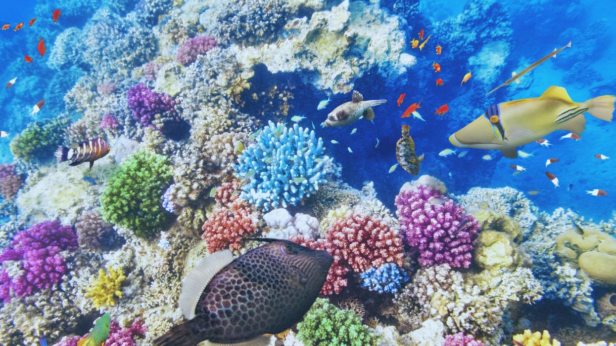
Underwater world with corals and tropical fish.
Coral reefs are usually relegated to bit player status in television and movies, providing splashes of background color for "Shark Week," "Finding Nemo," and other marine-based entertainment.
In real life, the reefs are an absolutely crucial component of the ecosystem for both oceans and land, rivaling only the rain forests in their biological complexity. They provide shelter and sustenance for up to a quarter of all marine life, oxygenate the water, help protect coastlines from erosion, and support thousands of tourism jobs and businesses.
Genetic engineering could help scientists rebuild the reefs that have been lost, and turn those still alive into a souped-up version that can withstand warmer and even more acidic waters.
But the warming of the world's oceans -- exacerbated by an El Nino event that occurred between 2014 and 2016 -- has been putting the world's reefs under tremendous pressure. Their vibrant colors are being replaced by sepulchral whites and tans.
That's the result of bleaching -- a phenomenon that occurs when the warming waters impact the efficiency of the algae that live within the corals in a symbiotic relationship, providing nourishment via photosynthesis and eliminating waste products. The corals will often "shuffle" their resident algae, reacting in much the same way a landlord does with a non-performing tenant -- evicting them in the hopes of finding a better resident. But when better-performing algae does not appear, the corals become malnourished, eventually becoming deprived of their color and then their lives.
The situation is dire: Two-thirds of Australia's Great Barrier Reef have undergone a bleaching event in recent years, and it's believed up to half of that reef has died.
Moreover, hard corals are the ocean's redwood trees. They take centuries to grow, meaning it could take centuries or more to replace them.
Recent developments in genetic engineering -- and an accidental discovery by researchers at a Florida aquarium -- provide opportunities for scientists to potentially rebuild a large proportion of the reefs that have been lost, and perhaps turn those still alive into a souped-up version that can withstand warmer and even more acidic waters. But many questions have yet to be answered about both the biological impact on the world's oceans, and the ethics of reengineering the linchpin of its ecosystem.
How did we get here?
Coral bleaching was a regular event in the oceans even before they began to warm. As a result, natural selection weeds out the weaker species, says Rachel Levin, an American-born scientist who has performed much of her graduate work in Australia. But the current water warming trend is happening at a much higher rate than it ever has in nature, and neither the coral nor the algae can keep up.
"There is a big concern about giving one variant a huge fitness advantage, have it take over and impact the natural variation that is critical in changing environments."
In a widely-read paper published last year in the journal Frontiers in Microbiology, Levin and her colleagues put forth a fairly radical notion for preserving the coral reefs: Genetically modify their resident algae.
Levin says the focus on algae is a pragmatic decision. Unlike coral, they reproduce extremely rapidly. In theory, a modified version could quickly inhabit and stabilize a reef. About 70 percent of algae -- all part of the genus symbiodinium -- are host generalists. That means they will insert themselves into any species of coral.
In recent years, work on mapping the genomes of both algae and coral has been progressing rapidly. Scientists at Stanford University have recently been manipulating coral genomes using larvae manipulated with the CRISPR/Cas9 technology, although the experimentation has mostly been limited to its fluorescence.
Genetically modifying the coral reefs could seem like a straightforward proposition, but complications are on the horizon. Levin notes that as many as 20 different species of algae can reside within a single coral, so selecting the best ones to tweak may pose a challenge.
"The entire genus is made up of thousands of subspecies, all very genetically distinct variants. There is a huge genetic diversity, and there is a big concern about giving one variant a huge fitness advantage, have it take over and impact the natural variation that is critical in changing environments," Levin says.
Genetic modifications to an algae's thermal tolerance also poses the risk of what Levin calls an "off-target effect." That means a change to one part of the genome could lead to changes in other genes, such as those regulating growth, reproduction, or other elements crucial to its relationship with coral.
Phillip Cleves, a postdoctoral researcher at Stanford who has participated in the CRISPR/Cas9 work, says that future research will focus on studying the genes in coral that regulate the relationship with the algae. But he is so concerned about the ethical issues of genetically manipulating coral to adapt to a changing climate that he declined to discuss it in detail. And most coral species have not yet had their genomes fully mapped, he notes, suggesting that such work could still take years.
An Alternative: Coral Micro-fragmentation
In the meantime, there is another technique for coral preservation led by David Vaughan, senior scientist and program manager at the Mote Marine Laboratory and Aquarium in Sarasota, Florida.
Vaughan's research team has been experimenting in the past decade with hard coral regeneration. Their work had been slow and painstaking, since growing larvae into a coral the size of a quarter takes three years.
The micro-fragmenting process in some ways raises fewer ethical questions than genetically altering the species.
But then, one day in 2006, Vaughan accidentally broke off a tiny piece of coral in the research aquarium. That fragment grew to the size of a quarter in three months, apparently the result of the coral's ability to rapidly regenerate when injured. Further research found that breaking coral in this manner -- even to the size of a single polyp -- led to rapid growth in more than two-dozen species.
Mote is using this process, known as micro-fragmentation, to grow large numbers of coral rapidly, often fusing them on top of larger pieces of dead coral. These coral heads are then planted in the Florida Keys, which has experienced bleaching events over 12 of the last 14 years. The process has sped up almost exponentially; Mote has planted some 36,000 pieces of coral to date, but Vaughan says it's on track to plant 35,000 more pieces this year alone. That sum represents between 20 to 30 acres of restored reef. Mote is on track to plant another 100,000 pieces next year.
This rapid reproduction technique in some ways allows Mote scientists to control for the swift changes in ocean temperature, acidification and other factors. For example, using surviving pieces of coral from areas that have undergone bleaching events means these hardier strains will propagate much faster than nature allows.
Vaughan recently visited the Yucatan Peninsula to work with Mexican researchers who are going to embark on a micro-fragmenting initiative of their own.
The micro-fragmenting process in some ways raises fewer ethical questions than genetically altering the species, although Levin notes that this could also lead to fewer varieties of corals on the ocean floor -- a potential flattening of the colorful backdrops seen in television and movies.
But Vaughan has few qualms, saying this is an ecological imperative. He suggests that micro-fragmentation could serve as a stopgap until genomic technologies further advance.
"We have to use the technology at hand," he says. "This is a lot like responding when a forest burns down. We don't ask questions. We plant trees."
A new type of cancer therapy is shrinking deadly brain tumors with just one treatment
MRI scans after a new kind of immunotherapy for brain cancer show remarkable progress in one patient just days after the first treatment.
Few cancers are deadlier than glioblastomas—aggressive and lethal tumors that originate in the brain or spinal cord. Five years after diagnosis, less than five percent of glioblastoma patients are still alive—and more often, glioblastoma patients live just 14 months on average after receiving a diagnosis.
But an ongoing clinical trial at Mass General Cancer Center is giving new hope to glioblastoma patients and their families. The trial, called INCIPIENT, is meant to evaluate the effects of a special type of immune cell, called CAR-T cells, on patients with recurrent glioblastoma.
How CAR-T cell therapy works
CAR-T cell therapy is a type of cancer treatment called immunotherapy, where doctors modify a patient’s own immune system specifically to find and destroy cancer cells. In CAR-T cell therapy, doctors extract the patient’s T-cells, which are immune system cells that help fight off disease—particularly cancer. These T-cells are harvested from the patient and then genetically modified in a lab to produce proteins on their surface called chimeric antigen receptors (thus becoming CAR-T cells), which makes them able to bind to a specific protein on the patient’s cancer cells. Once modified, these CAR-T cells are grown in the lab for several weeks so that they can multiply into an army of millions. When enough cells have been grown, these super-charged T-cells are infused back into the patient where they can then seek out cancer cells, bind to them, and destroy them. CAR-T cell therapies have been approved by the US Food and Drug Administration (FDA) to treat certain types of lymphomas and leukemias, as well as multiple myeloma, but haven’t been approved to treat glioblastomas—yet.
CAR-T cell therapies don’t always work against solid tumors, such as glioblastomas. Because solid tumors contain different kinds of cancer cells, some cells can evade the immune system’s detection even after CAR-T cell therapy, according to a press release from Massachusetts General Hospital. For the INCIPIENT trial, researchers modified the CAR-T cells even further in hopes of making them more effective against solid tumors. These second-generation CAR-T cells (called CARv3-TEAM-E T cells) contain special antibodies that attack EFGR, a protein expressed in the majority of glioblastoma tumors. Unlike other CAR-T cell therapies, these particular CAR-T cells were designed to be directly injected into the patient’s brain.
The INCIPIENT trial results
The INCIPIENT trial involved three patients who were enrolled in the study between March and July 2023. All three patients—a 72-year-old man, a 74-year-old man, and a 57-year-old woman—were treated with chemo and radiation and enrolled in the trial with CAR-T cells after their glioblastoma tumors came back.
The results, which were published earlier this year in the New England Journal of Medicine (NEJM), were called “rapid” and “dramatic” by doctors involved in the trial. After just a single infusion of the CAR-T cells, each patient experienced a significant reduction in their tumor sizes. Just two days after receiving the infusion, the glioblastoma tumor of the 72-year-old man decreased by nearly twenty percent. Just two months later the tumor had shrunk by an astonishing 60 percent, and the change was maintained for more than six months. The most dramatic result was in the 57-year-old female patient, whose tumor shrank nearly completely after just one infusion of the CAR-T cells.
The results of the INCIPIENT trial were unexpected and astonishing—but unfortunately, they were also temporary. For all three patients, the tumors eventually began to grow back regardless of the CAR-T cell infusions. According to the press release from MGH, the medical team is now considering treating each patient with multiple infusions or prefacing each treatment with chemotherapy to prolong the response.
While there is still “more to do,” says co-author of the study neuro-oncologist Dr. Elizabeth Gerstner, the results are still promising. If nothing else, these second-generation CAR-T cell infusions may someday be able to give patients more time than traditional treatments would allow.
“These results are exciting but they are also just the beginning,” says Dr. Marcela Maus, a doctor and professor of medicine at Mass General who was involved in the clinical trial. “They tell us that we are on the right track in pursuing a therapy that has the potential to change the outlook for this intractable disease.”
A recent study in The Lancet Oncology showed that AI found 20 percent more cancers on mammogram screens than radiologists alone.
Since the early 2000s, AI systems have eliminated more than 1.7 million jobs, and that number will only increase as AI improves. Some research estimates that by 2025, AI will eliminate more than 85 million jobs.
But for all the talk about job security, AI is also proving to be a powerful tool in healthcare—specifically, cancer detection. One recently published study has shown that, remarkably, artificial intelligence was able to detect 20 percent more cancers in imaging scans than radiologists alone.
Published in The Lancet Oncology, the study analyzed the scans of 80,000 Swedish women with a moderate hereditary risk of breast cancer who had undergone a mammogram between April 2021 and July 2022. Half of these scans were read by AI and then a radiologist to double-check the findings. The second group of scans was read by two researchers without the help of AI. (Currently, the standard of care across Europe is to have two radiologists analyze a scan before diagnosing a patient with breast cancer.)
The study showed that the AI group detected cancer in 6 out of every 1,000 scans, while the radiologists detected cancer in 5 per 1,000 scans. In other words, AI found 20 percent more cancers than the highly-trained radiologists.

But even though the AI was better able to pinpoint cancer on an image, it doesn’t mean radiologists will soon be out of a job. Dr. Laura Heacock, a breast radiologist at NYU, said in an interview with CNN that radiologists do much more than simply screening mammograms, and that even well-trained technology can make errors. “These tools work best when paired with highly-trained radiologists who make the final call on your mammogram. Think of it as a tool like a stethoscope for a cardiologist.”
AI is still an emerging technology, but more and more doctors are using them to detect different cancers. For example, researchers at MIT have developed a program called MIRAI, which looks at patterns in patient mammograms across a series of scans and uses an algorithm to model a patient's risk of developing breast cancer over time. The program was "trained" with more than 200,000 breast imaging scans from Massachusetts General Hospital and has been tested on over 100,000 women in different hospitals across the world. According to MIT, MIRAI "has been shown to be more accurate in predicting the risk for developing breast cancer in the short term (over a 3-year period) compared to traditional tools." It has also been able to detect breast cancer up to five years before a patient receives a diagnosis.
The challenges for cancer-detecting AI tools now is not just accuracy. AI tools are also being challenged to perform consistently well across different ages, races, and breast density profiles, particularly given the increased risks that different women face. For example, Black women are 42 percent more likely than white women to die from breast cancer, despite having nearly the same rates of breast cancer as white women. Recently, an FDA-approved AI device for screening breast cancer has come under fire for wrongly detecting cancer in Black patients significantly more often than white patients.
As AI technology improves, radiologists will be able to accurately scan a more diverse set of patients at a larger volume than ever before, potentially saving more lives than ever.

