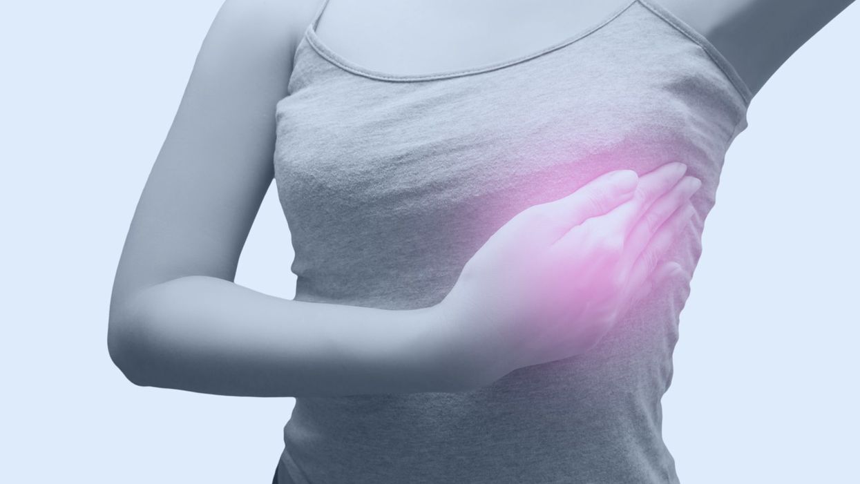
A woman checking her breast for the presence of concerning lumps.
Nancy Cappello was proactive. When she turned 36, she had a baseline mammogram, a standard medical recommendation in the late 1980s and early 1990s as a comparison tool for future screenings. At 40, Cappello started getting them annually.
Her breast surgeon estimated the cancer had been festering for four to five years under the radar of her annual mammograms.
Six weeks after her 11th-consecutive normal mammogram, she was diagnosed with Stage IIIc breast cancer.
A doctor felt a lump while doing a breast exam during her annual physical and a subsequent ultrasound detected cancer that had spread to 13 lymph nodes. That's when Cappello, then 51, learned she had dense breast tissue, making mammography less likely to detect tumors in her breasts.
She also discovered through her own research that she was among the 40 to 50 percent of women with dense breast tissue — almost half the female population — but medical protocol did not require physicians to inform women of their dense tissue status. If she had known, she said, she would have gotten an ultrasound every year in addition to a mammogram that could have detected the cancer much earlier. Cappello said her breast surgeon estimated the cancer had been festering for four to five years under the radar of her annual mammograms.
Although ultrasound as a cancer screening tool has been available for decades, technological advances are helping doctors find more invasive cancers in women with dense breasts, in turn giving women who know their tissue status the opportunity for earlier detection and treatment.
"We know that the gold standard for breast cancer screening is mammography, but in women with dense breast tissue, up to one third of breast cancers can be missed with this modality alone."
Dr. Georgia Giakoumis Spear, chief of the department of breast imaging at NorthShore University HealthSystem in suburban Chicago and assistant professor of radiology at the University of Chicago, has been a leader in developing standards for the use of new ultrasound technology. She is leading a study to develop more specific national guidelines around the use of Automated Whole Breast Tissue Ultrasound (ABUS), a non-invasive procedure in which sound waves are used to scan breast tissue while a patient lies on her back with her arm over her head.
Approved by the Food and Drug Administration in 2012, ABUS provides higher quality 3D images and faster delivery to provide more accurate results than past ultrasound technology. The scan does not involve radiation, and a practitioner can complete the process in 15 to 20 minutes, from patient preparation to image creation. NorthShore has been using ABUS since 2015, Dr. Spear said, and the technology can improve breast cancer detection in women with dense breasts by up to 55 percent.
"We know that the gold standard for breast cancer screening is mammography, but in women with dense breast tissue, up to one third of breast cancers can be missed with this modality alone," Spear says. "And when we supplement screening with ultrasound in this population of women, we have found a large number of cancers by ultrasound that are not visible on the mammogram."
Mammography should still be used as the first step for breast cancer detection, but if an initial mammogram shows that a patient has dense breast tissue, studies encourage discussion of additional screening with ultrasound.
On a mammogram, dense tissue appears white. So do cancerous masses, making them easy to miss.
A radiologist determines tissue density, according to the American College of Radiology's Breast Imaging Reporting and Data System (BI-RADS). "A" and "B" breast density categories designate ratios of mostly fatty, or non-dense tissues, while the "C" and "D" categories designate heterogeneously dense and extremely dense tissue, respectively. Such patients would be classified as having dense tissue. Younger women, women with lower levels of body fat and women undergoing hormone therapy are more likely to have C and D breast density.
On a mammogram, dense tissue appears white. So do cancerous masses, making them easy to miss. Fatty tissue, in comparison, appears black, making tumors easier to spot.
The FDA stated among its policy goals for 2018 that it's placing an improved focus on recognizing technological advances to help "ensure women get the most relevant, up-to-date information about their breast density, which is now recognized as a risk factor for breast cancer." An article in the March 2018 Journal of the American College of Radiology recommended supplemental screening for women with higher-than-average breast cancer risk, placing women with dense breast tissue in that category.
To be sure, some in the medical community are reluctant to push for ultrasounds, saying that a mammogram might be enough even if the woman has dense breast tissue. A patient is advised to discuss the option of ultrasound with her physician and they can decide from there.
Access to such information became political for Cappello after her diagnosis in 2004. She said that as she underwent six surgeries, a mastectomy, chemotherapy, radiation and hormone therapy, she asked doctors why they weren't required to inform women of their dense breast tissue status. Her dissatisfaction with their responses led to the formation of Are You Dense, Inc., an advocacy group aimed to inform women of their medical options while working to pass legislation mandating that women know their tissue status. Other legislation has focused on mandating insurance coverage for breast ultrasounds.

Nancy Cappello.
(Courtesy)
Cappello's work led Connecticut to become the first state to pass an information law in 2009, and 35 states now have similar requirements. Depending on the state, the law could mandate that certain language or information about breast density be included in the patient's mammogram results, or require physicians to tell women about dense tissue if their breast density falls in the BI-RADS categories C and D. Other states might require that patients be given general information about breast density and advice to discuss their options with a physician. (Note: There is a chart on Cappello's website that shows what laws exist – or don't – in each state.)
Through her site and social media, she's connected with other women who've lobbied for laws in their states, including Dr. Spear, who recently testified before legislative committees in Illinois as they considered companion bills. The Illinois legislation is expected to be signed into law this summer.
"There should be no excuses," Cappello says. "Women should have this information. There should be no concealing or hiding of her status."
Jamie Rettinger with his now fiance Amie Purnel-Davis, who helped him through the clinical trial.
Melanoma is the deadliest form of skin cancer. About 85,000 people are diagnosed with it each year in the U.S. and more than 8,000 die of the cancer when it spreads to other parts of the body, according to the Centers for Disease Control and Prevention (CDC).
There are two peaks in diagnosis of melanoma; one is in younger women ages 30-40 and often is tied to past use of tanning beds; the second is older men 60+ and is related to outdoor activity from farming to sports. Light-skinned people have a twenty-times greater risk of melanoma than do people with dark skin.
"When I graduated from medical school, in 2005, melanoma was a death sentence" --Diwakar Davar.
Jamie had a follow up PET scan about six months after his surgery. A suspicious spot on his lung led to a biopsy that came back positive for melanoma. The cancer had spread. Treatment with a monoclonal antibody (nivolumab/Opdivo®) didn't prove effective and he was referred to the UPMC Hillman Cancer Center in Pittsburgh, a four-hour drive from his home in western Ohio.
An alternative monoclonal antibody treatment brought on such bad side effects, diarrhea as often as 15 times a day, that it took more than a week of hospitalization to stabilize his condition. The only options left were experimental approaches in clinical trials.
Early research
"When I graduated from medical school, in 2005, melanoma was a death sentence" with a cure rate in the single digits, says Diwakar Davar, 39, an oncologist at UPMC Hillman Cancer Center who specializes in skin cancer. That began to change in 2010 with introduction of the first immunotherapies, monoclonal antibodies, to treat cancer. The antibodies attach to PD-1, a receptor on the surface of T cells of the immune system and on cancer cells. Antibody treatment boosted the melanoma cure rate to about 30 percent. The search was on to understand why some people responded to these drugs and others did not.
At the same time, there was a growing understanding of the role that bacteria in the gut, the gut microbiome, plays in helping to train and maintain the function of the body's various immune cells. Perhaps the bacteria also plays a role in shaping the immune response to cancer therapy.
One clue came from genetically identical mice. Animals ordered from different suppliers sometimes responded differently to the experiments being performed. That difference was traced to different compositions of their gut microbiome; transferring the microbiome from one animal to another in a process known as fecal transplant (FMT) could change their responses to disease or treatment.
When researchers looked at humans, they found that the patients who responded well to immunotherapies had a gut microbiome that looked like healthy normal folks, but patients who didn't respond had missing or reduced strains of bacteria.
Davar and his team knew that FMT had a very successful cure rate in treating the gut dysbiosis of Clostridioides difficile, a persistant intestinal infection, and they wondered if a fecal transplant from a patient who had responded well to cancer immunotherapy treatment might improve the cure rate of patients who did not originally respond to immunotherapies for melanoma.
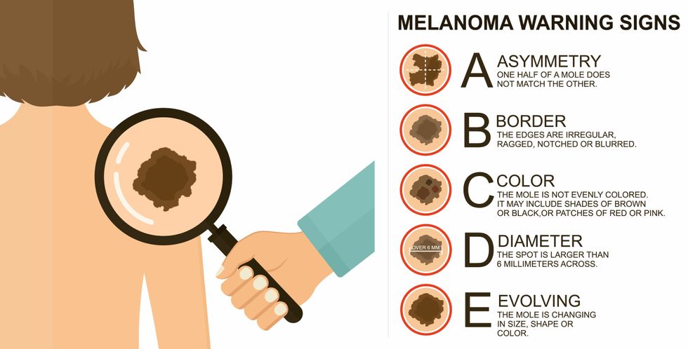
The ABCDE of melanoma detection
Adobe Stock
Clinical trial
"It was pretty weird, I was totally blasted away. Who had thought of this?" Jamie first thought when the hypothesis was explained to him. But Davar's explanation that the procedure might restore some of the beneficial bacterial his gut was lacking, convinced him to try. He quickly signed on in October 2018 to be the first person in the clinical trial.
Fecal donations go through the same safety procedures of screening for and inactivating diseases that are used in processing blood donations to make them safe for transfusion. The procedure itself uses a standard hollow colonoscope designed to screen for colon cancer and remove polyps. The transplant is inserted through the center of the flexible tube.
Most patients are sedated for procedures that use a colonoscope but Jamie doesn't respond to those drugs: "You can't knock me out. I was watching them on the TV going up my own butt. It was kind of unreal at that point," he says. "There were about twelve people in there watching because no one had seen this done before."
A test two weeks after the procedure showed that the FMT had engrafted and the once-missing bacteria were thriving in his gut. More importantly, his body was responding to another monoclonal antibody (pembrolizumab/Keytruda®) and signs of melanoma began to shrink. Every three months he made the four-hour drive from home to Pittsburgh for six rounds of treatment with the antibody drug.
"We were very, very lucky that the first patient had a great response," says Davar. "It allowed us to believe that even though we failed with the next six, we were on the right track. We just needed to tweak the [fecal] cocktail a little better" and enroll patients in the study who had less aggressive tumor growth and were likely to live long enough to complete the extensive rounds of therapy. Six of 15 patients responded positively in the pilot clinical trial that was published in the journal Science.
Davar believes they are beginning to understand the biological mechanisms of why some patients initially do not respond to immunotherapy but later can with a FMT. It is tied to the background level of inflammation produced by the interaction between the microbiome and the immune system. That paper is not yet published.
Surviving cancer
It has been almost a year since the last in his series of cancer treatments and Jamie has no measurable disease. He is cautiously optimistic that his cancer is not simply in remission but is gone for good. "I'm still scared every time I get my scans, because you don't know whether it is going to come back or not. And to realize that it is something that is totally out of my control."
"It was hard for me to regain trust" after being misdiagnosed and mistreated by several doctors he says. But his experience at Hillman helped to restore that trust "because they were interested in me, not just fixing the problem."
He is grateful for the support provided by family and friends over the last eight years. After a pause and a sigh, the ruggedly built 47-year-old says, "If everyone else was dead in my family, I probably wouldn't have been able to do it."
"I never hesitated to ask a question and I never hesitated to get a second opinion." But Jamie acknowledges the experience has made him more aware of the need for regular preventive medical care and a primary care physician. That person might have caught his melanoma at an earlier stage when it was easier to treat.
Davar continues to work on clinical studies to optimize this treatment approach. Perhaps down the road, screening the microbiome will be standard for melanoma and other cancers prior to using immunotherapies, and the FMT will be as simple as swallowing a handful of freeze-dried capsules off the shelf rather than through a colonoscopy. Earlier this year, the Food and Drug Administration approved the first oral fecal microbiota product for C. difficile, hopefully paving the way for more.
An older version of this hit article was first published on May 18, 2021