This Resistance Fighter Invented Dialysis in Nazi-Occupied Holland

When Willem Johan Kolff invented dialysis, the "father" of artificial organs was just getting started.
One of the Netherlands’ most famous pieces of pop culture is “Soldier of Orange.” It’s the title of the country’s most celebrated war memoir, movie and epic stage musical, all of which detail the exploits of the nation’s resistance fighters during World War II.
Willem Johan Kolff was a member of the Dutch resistance, but he doesn’t rate a mention in the “Solider of Orange” canon. Yet his wartime toils in a rural backwater not only changed medicine, but the world.
Kolff had been a physician less than two years before Germany invaded the Netherlands in May 1940. He had been engaged in post-graduate studies at the University of Gronigen but withdrew because he refused to accommodate the demands of the Nazi occupiers. Kolff’s Jewish supervisor made an even starker choice: He committed suicide.
After his departure from the university, Kolff took a job managing a small hospital in Kampen. Located 50 miles from the heavily populated coastal region, the facility was far enough away from the prying eyes of Germans that not only could Kolff care for patients, he could hide fellow resistance fighters and even Jewish refugees in relative safety. Kolff coached many of them to feign convincing terminal illnesses so the Nazis would allow them to remain in the hospital.
Despite the demands of practicing medicine and resistance work, Kolff still found time to conduct research. He had been haunted and inspired when, not long before the Nazi invasion, one of his patients died in agony from kidney disease. Kolff wanted to find a way to save future patients.
He broke his problem down to a simple task: If he could remove 20 grams of urea from a patient’s blood in 24 hours, they would survive. He began experimenting with ways to filter blood and return it to a patient’s body. Since the war had ground all non-military manufacturing to a halt, he was mostly forced to make do with material he could find at the hospital and around Kampen. Kolff eventually built a device from a washing machine parts, juice cans, sausage casings, a valve from an old Ford automobile radiator, and even scrap from a downed German aircraft.
The world’s first dialysis machine was hardly imposing; it resembled a rotating drum for a bingo game or raffle. Yet it carried on the highly sophisticated task of moving a patient’s blood through a semi-permeable membrane (about a 50-foot length of sausage casings) into a saline solution that drew out urea while leaving the blood cells untouched.
In emigrating to the U.S. to practice medicine, Kolff's intent was twofold: Advocate for a wider adoption of dialysis, and work on new projects. He wildly succeeded at both.
Kolff began using the machine to treat patients in 1943, most of whom had lapsed into comas due to their kidney failure. But like most groundbreaking medical devices, it was not an immediate success. By the end of the war, Kolff had dialyzed more than a dozen patients, but all had died. He briefly suspended use of the device after the Allied invasion of Europe, but he continued to refine its operation and the administration of blood thinners to patients.
In September 1945, Kolff dialyzed another comatose patient, 67-year-old Sofia Maria Schafstadt. She regained consciousness after 11 hours, and would live well into the 1950s with Kolff’s assistance. Yet this triumph contained a dark irony: At the time of her treatment, Schafstadt had been imprisoned for collaborating with the Germans.
With a tattered Europe struggling to overcome the destruction of the war, Kolff and his family emigrated to the U.S. in 1950, where he began working for the Cleveland Clinic while undergoing the naturalization process so he could practice medicine in the U.S. His intent was twofold: Advocate for a wider adoption of dialysis, and work on new projects. He wildly succeeded at both.
By the mid-1950s, dialysis machines had become reliable and life-saving medical devices, and Kolff had become a U.S. citizen. About that time he invented a membrane oxygenator that could be used in heart bypass surgeries. This was a critical component of the heart-lung machine, which would make heart transplants possible and bypass surgeries routine. He also invented among the very first practical artificial hearts, which in 1957 kept a dog alive for 90 minutes.
Kolff moved to the University of Utah in 1967 to become director of its Institute for Biomedical Engineering. It was a promising time for such a move, as the first successful transplant of a donor heart to a human occurred that year. But he was interested in going a step further and creating an artificial heart for human use.
It took more than a decade of tinkering and research, but in 1982, a team of physicians and engineers led by Kolff succeeded in implanting the first artificial heart in dentist Barney Clark, whose failing health disqualified him from a heart transplant. Although Clark died in March 1983 after 112 days tethered to the device, that it kept him alive generated international headlines. While graduate student Robert Jarvik received the named credit for the heart, he was directly supervised by Kolff, whose various endeavors into artificial organ research at the University of Utah were segmented into numerous teams.
Forty years later, several artificial hearts have been approved for use by the Food and Drug Administration, although all are a “bridge” that allow patients to wait for a transplant.
Kolff continued researching and tinkering with biomedical devices – including artificial eyes and ears – until he retired in 1997 at the age of 86. When he died in 2009, the medical community acknowledged that he was not only a pioneer in biotechnology, but the “father” of artificial organs.
How Genetic Testing and Targeted Treatments Are Helping More Cancer Patients Survive
Scientists are studying cancer genomes to more precisely diagnose their patients' diseases - offering hope for targeted drug treatments.
Late in 2018, Chris Reiner found himself “chasing a persistent cough” to figure out a cause. He talked to doctors; he endured various tests, including an X-ray. Initially, his physician suspected bronchitis. After several months, he still felt no improvement. In May 2019, his general practitioner recommended that Reiner, a business development specialist for a Seattle-based software company, schedule a CAT scan.
Reiner knew immediately that his doctor asking him to visit his office to discuss the results wasn’t a good sign. The longtime resident of Newburyport, MA, remembers dreading “that conversation that people who learn they have cancer have.”
“The doctor handed me something to look at, and the only thing I remember after that was everything went blank all around me,” Reiner, 50, reveals. “It was the magnitude of what he was telling me, that I had a malignant mass in my lung.”
Next, he recalls, he felt ushered into “the jaws of the medical system very quickly.” He spent a couple of days meeting with a team of doctors at Beth Israel Deaconess Medical Center in nearby Boston. One of them was from a medical field he hadn’t even known existed, a pulmonary interventionist, who would perform a biopsy on the mass in his lung.
“Knowing there was a medicine for my particular type of cancer was like a weight lifted off my shoulders."
A week later he and his wife Allison returned to meet with the oncologist, radiologist, pulmonary interventionist – his medical team. They confirmed his initial diagnosis: Stage 4 metastatic lung cancer that had spread to several parts of his body. “We just sat there, stunned,” he says. “I felt like I was getting hit by a wrecking ball over and over.”
An onslaught of medical terminology about what they had identified flowed over the shocked couple, but then the medical team switched gears, he recalls. They offered hope. “They told me, ‘Hey, you’re not a smoker, so that’s good,’” Reiner says. “‘There’s a good chance that what’s driving this disease for you is actually a genetic mutation, and we have ways to understand more about what that could be through some simple testing.’”
They told him about Foundation Medicine, a company launched in neighboring Cambridge, MA, in 2009 that develops, manufactures, and sells genomic profiling assays. These are tests that, according to the company’s website, “can analyze a broad panel of genes to detect the four main classes of genomic alterations known to drive cancer growth.” With these insights, certain patients can be matched with therapies targeted specifically for the genetic driver(s) of their cancer. The company maintains one of the largest cancer genomic databases in the world, with more than 500,000 patient samples profiled, and they have more than 65 biopharma partners.
According to Foundation Medicine, they are the only company that has FDA-approved tests for both tissue- and blood-based comprehensive genomic profiling tests. One other company has an FDA-approved biopsy test, and several other companies offer tissue-based genomic profiling. Additionally, several major cancer centers like Memorial Sloan Kettering in New York and Anderson Cancer Center in Texas have their own such testing platforms.
Currently, genomic profiling is more accessible for patients with advanced cancer, due to broader insurance coverage in later stages of disease.
“Right now, the vast majority of patients either have cancers for which we don’t have treatments or they have genetic alterations that are not known,” says Jorge Garcia, MD, Division Chief, Solid Tumor Oncology, UH Cleveland Medical Center, which has its own CGP testing platform. “However, a significant proportion of patients with advanced cancer have alterations that we can tap for therapeutic purposes.”
Foundation Medicine estimates that in 2017, just over 5 percent of advanced solid cancer patients in the U.S. received CGP testing. In 2021, they estimate that number is between 25 to 30 percent of advanced solid cancer patients in the U.S., which doesn’t include patients who are tested with small (less than 50 genes) panels. Their panel tests for more than 300 cancer-related genes.

“The good news is the platforms we are developing are better and more comprehensive, and they’re going to continue to be larger data sets,” Dr. Garcia adds.
In Reiner’s case, his team ordered comprehensive genetic profiling on both his tissue and blood, from Foundation Medicine.
At this point, Reiner still wasn’t sure what genetic mutations were or how they factored into cancer or what comprehensive genomic profiling entailed. That day, though, his team ushered the Reiners into the world of precision oncology that placed him on much more sure footing to learn about and fight the specific lung cancer that had been troubling him for more than a year.
What genetic alterations were driving his cancer? Foundation Medicine’s tests were about to find out.
At the core of these tests is next generation sequencing, a DNA sequencing technology. Since 2009, this has revolutionized genomic research, according to the National Center for Biotechnology Information, because it allows an entire human genome to be sequenced within one day. Cancer genomics posits that cancer is caused by mutations and is a disease of the genome. Now, cancer genomes can be systemically studied in their entirety. For cancer patients such as Reiner, NGS can provide a more precise diagnosis and classification of the disease, more accurate prognosis, and potentially the identification of targeted drug treatments. Ultimately, the technology can provide the basis of personalized cancer management.
The detailed reports supply patients and their oncologists with extensive information about the patient’s genomic profile and potential treatment options that they can discuss together. Reiner trusted his doctors that this approach was worth the two- or three-week wait to receive the Foundation Medicine report and the specifically targeted treatment, rather than immediately jump into a round of chemotherapy. He is especially grateful now, he says, because the report delivered a great deal of relief from his previously exhausting and growing anxiety about having cancer.
Reiner and his team learned his lung cancer contained the epidermal growth factor receptor (EGFR) mutation. That biomarker enabled his oncologist to prescribe Tagrisso (osimertinib), a medication developed to directly target that genetic mutation.
“Knowing there was a medicine for my particular type of cancer was like a weight lifted off my shoulders,” he says. “It only took a week or two before my cough finally started subsiding. This pill goes right after the particular piece of genetic material in the tumor that’s causing its growth.”
Dr. Jerry Mitchell, director field medical oncology, Foundation Medicine, in Columbus, Ohio, explains that genomic profiling is generating substantial impacts today. “This is a technology that is the standard of care across many advanced malignancies that takes patients from chemotherapy-only options to very targeted options or immunotherapy options,” he says. “You can also look at complex biomarkers, and these are not specific genetic changes but different genes across the tumor to get a biomarker.”
According to Dr. Mitchell, Foundation Medicine’s technology can test more than 324 different cancer-related genes in a single test. Thus, a growing number of patients are benefitting from comprehensive genetic profiling, due to the rapidly growing number of targeted therapies. While not all of the cancers are treatable yet, the company uses that information to partner with researchers to find new potential therapies for patient groups that may have rare mutations.
Since his tumor’s diagnosis, Reiner has undergone chemotherapy and a couple surgeries to treat the metastatic cancer in other parts of his body, but the drug Tagrisso has significantly reduced his lung tumor. Now, having learned so much during the past couple of years, he is grateful for precision oncology. He still reflects on the probability that, had the Tagrisso pill not been available in May 2019, he might have only survived for another six months or a year.
“Comprehensive Genomic Profiling is not some future state, but in both the U.S. and Europe, it is a very standard, accepted, and recommended first step to knowing how to treat your cancer,” says Dr. Mitchell, adding that he feels fortunate to be an oncologist in this era. “However, we know there are still people not getting this recommended testing, so we still have opportunities to find many more patients and impact them by knowing the molecular profile of their cancer.”
The Cellular Secrets of “Young Blood” Are Starting to Be Unlocked
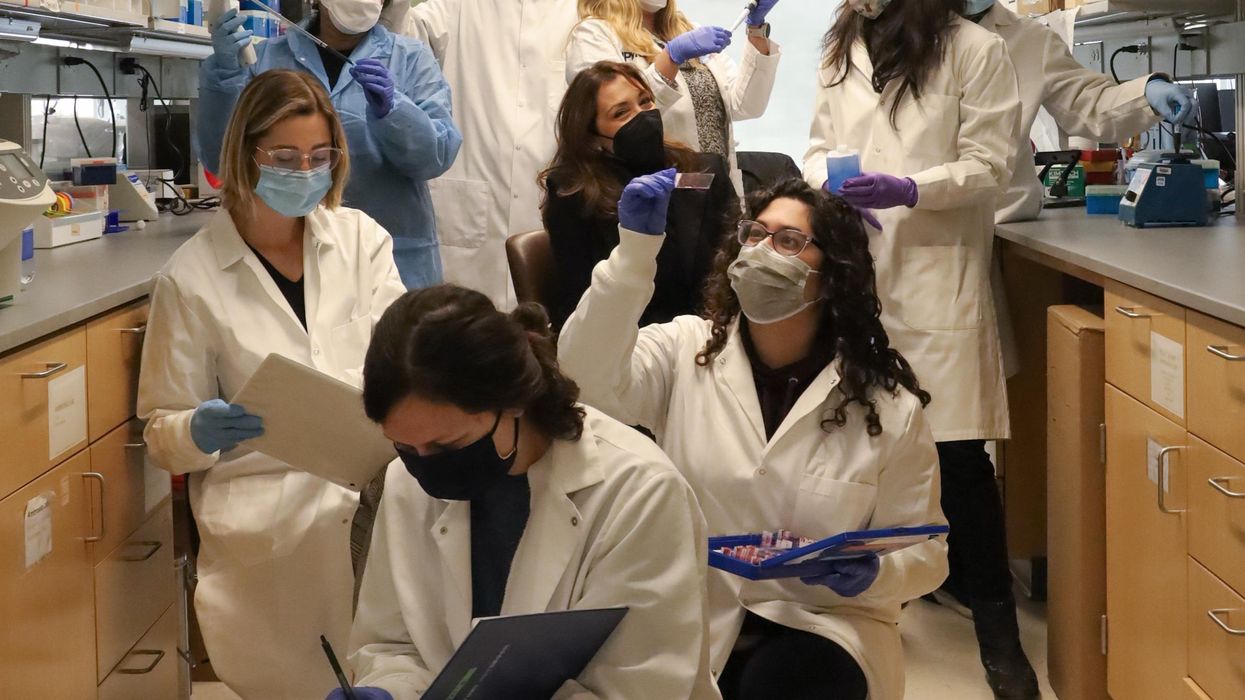
Fabrisia Ambrosio (center) surrounded by her lab staff.
The quest for an elixir to restore youthful health and vigor is common to most cultures and has prompted much scientific research. About a decade ago, Stanford scientists stitched together the blood circulatory systems of old and young mice in a practice called parabiosis. It seemed to rejuvenate the aged animals and spawned vampirish urban legends of Hollywood luminaries and tech billionaires paying big bucks for healthy young blood to put into their own aging arteries in the hope of reversing or at least forestalling the aging process.
It was “kind of creepy” and also inspiring to Fabrisia Ambrosio, then thousands of miles away and near the start of her own research career into the processes of aging. Her lab is at the University of Pittsburgh but on this cold January morning I am speaking with her via Zoom as she visits with family near her native Sao Paulo, Brazil. A gleaming white high rise condo and a lush tropical jungle split the view behind her, and the summer beach is just a few blocks away.
Ambrosio possesses the joy of a kid on Christmas morning who can't wait to see what’s inside the wrapping. “I’ve always had a love for research, my father was a physicist," she says, but interest in the human body pulled her toward biology as her education progressed in the U.S. and Canada.
Back in Pittsburgh, her lab first extended the work of others in aging by using the simpler process of injecting young blood into the tail vein of old mice and found that the skeletal muscles of the animals “displayed an enhanced capacity to regenerate.” But what was causing this improvement?
When Ambrosio injected old mice with young blood depleted of EVs, the regenerative effect practically disappeared.
The next step was to remove the extracellular vesicles (EVs) from blood. EVs are small particles of cells composed of a membrane and often a cargo inside that lipid envelope. Initially many scientists thought that EVs were simply taking out the garbage that cells no longer needed, but they would learn that one cell's trash could be another cell's treasure.
Metabolites, mRNA, and myriad other signaling molecules inside the EV can function as a complex network by which cells communicate with others both near and far. These cargoes can up and down-regulate gene expression, affecting cell activity and potentially the entire body. EVs are present in humans, the bacteria that live in and on us, even in plants; they likely communicate across all forms of life.
Being inside the EV membrane protects cargo from enzymes and other factors in the blood that can degrade it, says Kenneth Witwer, a researcher at Johns Hopkins University and program chair of the International Society for Extracellular Vesicles. The receptors on the surface of the EV provide clues to the type of cell from which it originated and the cell receptors to which it might later bind and affect.
When Ambrosio injected old mice with young blood depleted of EVs, the regenerative effect practically disappeared; purified EVs alone were enough to do the job. The team also looked at muscle cell gene expression after injections of saline, young blood, and EV-depleted young blood and found significant differences. She believes this means that the major effect of enhanced regenerative capacity was coming from the EVs, though free floating proteins within the blood may also contribute something to the effect.
One such protein, called klotho, is of great interest to researchers studying aging. The name was borrowed from the Fates of Greek mythology, which consists of three sisters; Klotho spins the thread of life that her sisters measure and cut. Ambrosio had earlier shown that supplementing klotho could enhance regenerative capacity in old animals. But as with most proteins, klotho is fragile, rapidly degrading in body fluids, or when frozen and thawed. She suspected that klotho could survive better as cargo enclosed within the membrane of an EV and shielded from degradation.
So she went looking for klotho inside the EVs they had isolated. Advanced imaging technology revealed that young EVs contained abundant levels of klotho mRNAs, but the number of those proteins was much lower in EVs from old mice. Ambrosio wrote in her most recent paper, published in December in Nature Aging. She also found that the stressors associated with aging reduced the communications capacity of EVs in muscle tissue and that could be only partially restored with young blood.
Researchers still don't understand how klotho functions at the cellular level, but they may not need to know that. Perhaps learning how to increase its production, or using synthetic biology to generate more copies of klotho mRNA, or adding cell receptors to better direct EVs to specific aging tissue will be sufficient to reap the anti-aging benefits.
“Very, very preliminary data from our lab has demonstrated that exercise may be altering klotho transcripts within aged extracellular vesicles" for the better Ambrosio teases. But we already know that exercise is good for us; understanding the cellular mechanism behind that isn't likely to provide additional motivation to get up off the couch. Many of us want a prescription, a pill that is easy to take, to slow our aging.
Ambrosio hopes that others will build upon the basic research from her lab, and that pharmaceutical companies will be able to translate and develop it into products that can pass through FDA review and help ameliorate the diseases of aging.

