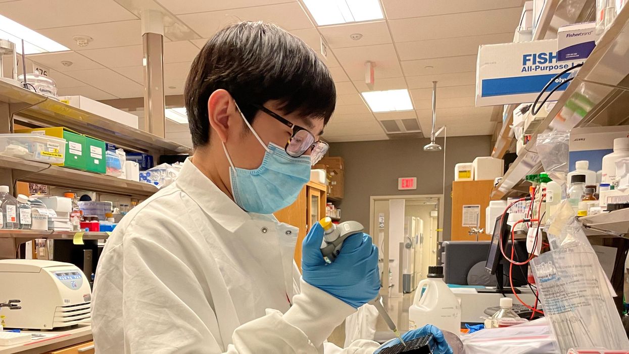
A new method could help the smallest of medicines hit their targets

Jacob Brenner and his partners at the University of Pennsylvania's Perelman School of Medicine are finding new ways to get nanomedicines to arrive at their targets.
Its strength is in its lack of size.
Using materials on the minuscule scale of nanometers (billionths of a meter), nanomedicines have the ability to provide treatment more precise than any other form of medicine. Under optimal circumstances, they can target specific cells and perform feats like altering the expression of proteins in tumors so that the tumors shrink.
Another appealing concept about nanomedicine is that treatment on a nano-scale, which is smaller yet than individual cells, can greatly decrease exposure to parts of the body outside the target area, thereby mitigating side effects.
But this young field's huge potential has met with an ongoing obstacle: the recipient's immune system tends to regard incoming nanomedicines as a threat and launches a complement protein attack. These complement proteins, which act together through a wave of reactions to get rid of troubling microorganisms, have had more than 500 million years to refine their craft, so they are highly effective.
Seeking to overcome a half-billion-year disadvantage, nanomaterials engineers have tried such strategies as creating so-called stealth nanoparticles.
“All new technologies face technical barriers, and it is the job of innovators to engineer solutions to them,” Brenner says.
Despite these clever attempts, nanomedicines largely keep failing to arrive at their intended destinations. According to the most comprehensive meta-analysis of nanomedicines in oncology, fewer than 1 percent of nanoparticles manage to reach their targets. The remaining 99-plus percent are expelled to the liver, spleen, or lungs – thereby squandering their therapeutic potential. Though these numbers seem discouraging, systems biologist Jacob Brenner remains undaunted. “All new technologies face technical barriers, and it is the job of innovators to engineer solutions to them,” he says.
Brenner and his fellow researchers at the Perelman School of Medicine at the University of Pennsylvania have recently devised a method that, in a study published in late 2021 involving sepsis-afflicted mice, saw a longer half-life of nanoparticles in the bloodstream. This effect is crucial because “the longer our nanoparticles circulate, the more time they have to reach their target organs,” says Brenner, the study's co-principal investigator. He works as a critical care physician at the Hospital of the University of Pennsylvania, where he also serves as an assistant professor of medicine.
The method used by Brenner's lab involves coating nanoparticles with natural suppressors that safeguard against a complement attack from the recipient's immune system. For this idea, he credits bacteria. “They are so much smarter than us,” he says.
Brenner points out that many species of bacteria have learned to coat themselves in a natural complement suppressor known as Factor H in order to protect against a complement attack.
Humans also have Factor H, along with an additional suppressor called Factor I, both of which flow through our blood. These natural suppressors “are recruited to the surface of our own cells to prevent complement [proteins] from attacking our own cells,” says Brenner.
Coating nanoparticles with a natural suppressor is a “very creative approach that can help tone and improve the activity of nanotechnology medicines inside the body,” says Avi Schroeder, an associate professor at Technion - Israel Institute of Technology, where he also serves as Head of the Targeted Drug Delivery and Personalized Medicine Group.
Schroeder explains that “being able to tone [down] the immune response to nanoparticles enhances their circulation time and improves their targeting capacity to diseased organs inside the body.” He adds how the approach taken by the Penn Med researchers “shows that tailoring the surface of the nanoparticles can help control the interactions the nanoparticles undergo in the body, allowing wider and more accurate therapeutic activity.”
Brenner says he and his research team are “working on the engineering details” to streamline the process. Such improvements could further subdue the complement protein attacks which for decades have proven the bane of nanomedical engineers.
Though these attacks have limited nanomedicine's effectiveness, the field has managed some noteworthy successes, such as the chemotherapy drugs Abraxane and Doxil, the first FDA-approved nanomedicine.
And amid the COVID-19 pandemic, nanomedicines became almost universally relevant with the vast circulation of the Moderna and Pfizer-BioNTech vaccines, both of which consist of lipid nanoparticles. “Without the nanoparticle, the mRNA would not enter the cells effectively and would not carry out the therapeutic goal,” Schroeder explains.
These vaccines, though, are “just the start of the potential transformation that nanomedicine will bring to the world,” says Brenner. He relates how nanomedicine is “joining forces with a number of other technological innovations,” such as cell therapies in which nanoparticles aim to reprogram T-cells to attack cancer.
With a similar degree of optimism, Schroeder says, “We will see further growing impact of nanotechnologies in the clinic, mainly by enabling gene therapy for treating and even curing diseases that were incurable in the past.”
Brenner says that in the next 10 to 15 years, “nanomedicine is likely to impact patients” contending with a “huge diversity” of conditions. “I can't wait to see how it plays out.”
A new type of cancer therapy is shrinking deadly brain tumors with just one treatment
MRI scans after a new kind of immunotherapy for brain cancer show remarkable progress in one patient just days after the first treatment.
Few cancers are deadlier than glioblastomas—aggressive and lethal tumors that originate in the brain or spinal cord. Five years after diagnosis, less than five percent of glioblastoma patients are still alive—and more often, glioblastoma patients live just 14 months on average after receiving a diagnosis.
But an ongoing clinical trial at Mass General Cancer Center is giving new hope to glioblastoma patients and their families. The trial, called INCIPIENT, is meant to evaluate the effects of a special type of immune cell, called CAR-T cells, on patients with recurrent glioblastoma.
How CAR-T cell therapy works
CAR-T cell therapy is a type of cancer treatment called immunotherapy, where doctors modify a patient’s own immune system specifically to find and destroy cancer cells. In CAR-T cell therapy, doctors extract the patient’s T-cells, which are immune system cells that help fight off disease—particularly cancer. These T-cells are harvested from the patient and then genetically modified in a lab to produce proteins on their surface called chimeric antigen receptors (thus becoming CAR-T cells), which makes them able to bind to a specific protein on the patient’s cancer cells. Once modified, these CAR-T cells are grown in the lab for several weeks so that they can multiply into an army of millions. When enough cells have been grown, these super-charged T-cells are infused back into the patient where they can then seek out cancer cells, bind to them, and destroy them. CAR-T cell therapies have been approved by the US Food and Drug Administration (FDA) to treat certain types of lymphomas and leukemias, as well as multiple myeloma, but haven’t been approved to treat glioblastomas—yet.
CAR-T cell therapies don’t always work against solid tumors, such as glioblastomas. Because solid tumors contain different kinds of cancer cells, some cells can evade the immune system’s detection even after CAR-T cell therapy, according to a press release from Massachusetts General Hospital. For the INCIPIENT trial, researchers modified the CAR-T cells even further in hopes of making them more effective against solid tumors. These second-generation CAR-T cells (called CARv3-TEAM-E T cells) contain special antibodies that attack EFGR, a protein expressed in the majority of glioblastoma tumors. Unlike other CAR-T cell therapies, these particular CAR-T cells were designed to be directly injected into the patient’s brain.
The INCIPIENT trial results
The INCIPIENT trial involved three patients who were enrolled in the study between March and July 2023. All three patients—a 72-year-old man, a 74-year-old man, and a 57-year-old woman—were treated with chemo and radiation and enrolled in the trial with CAR-T cells after their glioblastoma tumors came back.
The results, which were published earlier this year in the New England Journal of Medicine (NEJM), were called “rapid” and “dramatic” by doctors involved in the trial. After just a single infusion of the CAR-T cells, each patient experienced a significant reduction in their tumor sizes. Just two days after receiving the infusion, the glioblastoma tumor of the 72-year-old man decreased by nearly twenty percent. Just two months later the tumor had shrunk by an astonishing 60 percent, and the change was maintained for more than six months. The most dramatic result was in the 57-year-old female patient, whose tumor shrank nearly completely after just one infusion of the CAR-T cells.
The results of the INCIPIENT trial were unexpected and astonishing—but unfortunately, they were also temporary. For all three patients, the tumors eventually began to grow back regardless of the CAR-T cell infusions. According to the press release from MGH, the medical team is now considering treating each patient with multiple infusions or prefacing each treatment with chemotherapy to prolong the response.
While there is still “more to do,” says co-author of the study neuro-oncologist Dr. Elizabeth Gerstner, the results are still promising. If nothing else, these second-generation CAR-T cell infusions may someday be able to give patients more time than traditional treatments would allow.
“These results are exciting but they are also just the beginning,” says Dr. Marcela Maus, a doctor and professor of medicine at Mass General who was involved in the clinical trial. “They tell us that we are on the right track in pursuing a therapy that has the potential to change the outlook for this intractable disease.”
A recent study in The Lancet Oncology showed that AI found 20 percent more cancers on mammogram screens than radiologists alone.
Since the early 2000s, AI systems have eliminated more than 1.7 million jobs, and that number will only increase as AI improves. Some research estimates that by 2025, AI will eliminate more than 85 million jobs.
But for all the talk about job security, AI is also proving to be a powerful tool in healthcare—specifically, cancer detection. One recently published study has shown that, remarkably, artificial intelligence was able to detect 20 percent more cancers in imaging scans than radiologists alone.
Published in The Lancet Oncology, the study analyzed the scans of 80,000 Swedish women with a moderate hereditary risk of breast cancer who had undergone a mammogram between April 2021 and July 2022. Half of these scans were read by AI and then a radiologist to double-check the findings. The second group of scans was read by two researchers without the help of AI. (Currently, the standard of care across Europe is to have two radiologists analyze a scan before diagnosing a patient with breast cancer.)
The study showed that the AI group detected cancer in 6 out of every 1,000 scans, while the radiologists detected cancer in 5 per 1,000 scans. In other words, AI found 20 percent more cancers than the highly-trained radiologists.

But even though the AI was better able to pinpoint cancer on an image, it doesn’t mean radiologists will soon be out of a job. Dr. Laura Heacock, a breast radiologist at NYU, said in an interview with CNN that radiologists do much more than simply screening mammograms, and that even well-trained technology can make errors. “These tools work best when paired with highly-trained radiologists who make the final call on your mammogram. Think of it as a tool like a stethoscope for a cardiologist.”
AI is still an emerging technology, but more and more doctors are using them to detect different cancers. For example, researchers at MIT have developed a program called MIRAI, which looks at patterns in patient mammograms across a series of scans and uses an algorithm to model a patient's risk of developing breast cancer over time. The program was "trained" with more than 200,000 breast imaging scans from Massachusetts General Hospital and has been tested on over 100,000 women in different hospitals across the world. According to MIT, MIRAI "has been shown to be more accurate in predicting the risk for developing breast cancer in the short term (over a 3-year period) compared to traditional tools." It has also been able to detect breast cancer up to five years before a patient receives a diagnosis.
The challenges for cancer-detecting AI tools now is not just accuracy. AI tools are also being challenged to perform consistently well across different ages, races, and breast density profiles, particularly given the increased risks that different women face. For example, Black women are 42 percent more likely than white women to die from breast cancer, despite having nearly the same rates of breast cancer as white women. Recently, an FDA-approved AI device for screening breast cancer has come under fire for wrongly detecting cancer in Black patients significantly more often than white patients.
As AI technology improves, radiologists will be able to accurately scan a more diverse set of patients at a larger volume than ever before, potentially saving more lives than ever.

