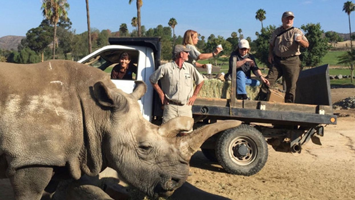
The Northern white rhinoceros Nola, the last one in the U.S. at that time in 2015, pictured here with author Jeanne Loring and Oliver Ryder (in truck), with a film crew and keepers in the San Diego Zoo's savanna. Nola sadly passed away that year.
I am a stem cell scientist. In my day job I work on developing ways to use stem cells to treat neurological disease – human disease. This is the story about how I became part of a group dedicated to rescuing the northern white rhinoceros from extinction.
The earth is now in an era that is called the "sixth mass extinction." The first extinction, 400 million years ago, put an end to 86 percent of the existing species, including most of the trilobites. When the earth grew hotter, dustier, or darker, it lost fish, amphibians, reptiles, plants, dinosaurs, mammals and birds. Each extinction event wiped out 80 to 90 percent of the life on the planet at the time. The first 5 mass extinctions were caused by natural disasters: volcanoes, fires, a meteor. But humans can take credit for the 6th.
Because of human activities that destroy habitats, creatures are now becoming extinct at a rate that is higher than any previously experienced. Some animals, like the giant panda and the California condor, have been pulled back from the brink of extinction by conserving their habitats, breeding in captivity, and educating the public about their plight.
But not the northern white rhino. This gentle giant is a vegetarian that can weigh up to 5,000 pounds. The rhino's weakness is its horn, which has become a valuable commodity because of the mistaken idea that it grants power and has medicinal value. Horns are not medicine; the horns are made of keratin, the same protein that is in fingernails. But as recently as 2017 more than 1,000 rhinos were slaughtered each year to harvest their horns.
All 6 rhino species are endangered. But the northern white has been devastated. Only two members of this species are alive now: Najin, age 32, and her daughter Fatu, 21, live in a protected park in Kenya. They are social animals and would prefer the company of other rhinos of their kind; but they can't know that they are the last two survivors of their entire species. No males exist anymore. The last male, Sudan, died in 2018 at age 45.
We are celebrating a huge milestone in the efforts to use stem cells to rescue the rhino.
I became involved in the rhino rescue project on a sunny day in February, 2008 at the San Diego Wild Animal Park in Escondido, about 30 miles north of my lab in La Jolla. My lab had relocated a couple of months earlier to Scripps Research Institute to start the Center for Regenerative Medicine for human stem cell research. To thank my staff for their hard work, I wanted to arrange a special treat. I contacted my friend Oliver Ryder, who is director of the Institute for Conservation Research at the zoo, to see if I could take them on a safari, a tour in a truck through the savanna habitat at the park.
This was the first of the "stem cell safaris" that the lab would enjoy over the next few years. On the safari we saw elands and cape buffalo, and fed giraffes and rhinos. And we talked about stem cells; in particular, we discussed a surprising technological breakthrough recently reported by the Japanese scientist Shinya Yamanaka that enabled conversion of ordinary skin cells into pluripotent stem cells.
Pluripotent stem cells can develop into virtually any cell type in the body. They exist when we are very young embryos; five days after we were just fertilized eggs, we became blastocysts, invisible tiny balls of a few hundred cells packed with the power to develop into an entire human being. Long before we are born, these cells of vast potential transform into highly specialized cells that generate our brains, our hearts, and everything else.
Human pluripotent stem cells from blastocysts can be cultured in the lab, and are called embryonic stem cells. But thanks to Dr. Yamanaka, anyone can have their skin cells reprogrammed into pluripotent stem cells, just like the ones we had when we were embryos. Dr. Yamanaka won the Nobel Prize for these cells, called "induced pluripotent stem cells" (iPSCs) several years later.
On our safari we realized that if we could make these reprogrammed stem cells from human skin cells, why couldn't we make them from animals' cells? How about endangered animals? Could such stem cells be made from animals whose skin cells had been being preserved since the 1970s in the San Diego Zoo's Frozen Zoo®? Our safari leader, Oliver Ryder, was the curator of the Frozen Zoo and knew what animal cells were stored in its giant liquid nitrogen tanks at −196°C (-320° F). The Frozen Zoo was established by Dr. Kurt Benirschke in 1975 in the hope that someday the collection would aid in rescue of animals that were on the brink of extinction. The frozen collection reached 10,000 cell lines this year.
We returned to the lab after the safari, and I asked my scientists if any of them would like to take on the challenge of making reprogrammed stem cells from endangered species. My new postdoctoral fellow, Inbar Friedrich Ben-Nun, raised her hand. Inbar had arrived only a few weeks earlier from Israel, and she was excited about doing something that had never been done before. Oliver picked the animals we would use. He chose his favorite animal, the critically endangered northern white rhinoceros, and the drill, which is an endangered primate related to the mandrill monkey,
When Inbar started work on reprogramming cells from the Frozen Zoo, there were 8 living northern rhinoceros around the world: Nola, Angalifu, Nesari, Nabire, Suni, Sudan, Najin, and Fatu. We chose to reprogram Fatu, the youngest of the remaining animals.
Through sheer determination and trial and error, Inbar got the reprogramming technique to work, and in 2011 we published the first report of iPSCs from endangered species in the scientific journal Nature Methods. The cover of the journal featured a drawing of an ark packed with animals that might someday be rescued through iPSC technology. By 2011, one of the 8 rhinos, Nesari, had died.
This kernel of hope for using iPSCs to rescue rhinos grew over the next 10 years. The zoo built the Rhino Rescue Center, and brought in 6 females of the closely related species, the southern white rhinoceros, from Africa. Southern white rhino populations are on the rise, and it appears that this species will survive, at least in captivity. The females are destined to be surrogate mothers for embryos made from northern white rhino cells, when eventually we hope to generate sperm and eggs from the reprogrammed stem cells, and fertilize the eggs in vitro, much the same as human IVF.

The author, Jeanne Loring, at the Rhino Rescue Center with one of the southern white rhino surrogates.
David Barker
As this project has progressed, we've been saddened by the loss of all but the last two remaining members of the species. Nola, the last northern white rhino in the U.S., who was at the San Diego Zoo, died in 2015.
But we are celebrating a huge milestone in the efforts to use stem cells to rescue the rhino. Just over a month ago, we reported that by reprogramming cells preserved in the Frozen Zoo, we produced iPSCs from stored cells of 9 northern white rhinos: Fatu, Najin, Nola, Suni, Nadi, Dinka, Nasima, Saut, and Angalifu. We also reprogrammed cells from two of the southern white females, Amani and Wallis.
We don't know when it will be possible to make a northern white rhino embryo; we have to figure out how to use methods already developed for laboratory mice to generate sperm and eggs from these cells. The male rhino Angalifu died in 2014, but ever since I saw beating heart cells derived from his very own cells in a culture dish, I've felt hope that he will one day have children who will seed a thriving new herd of northern white rhinos.
A new test called the EyeBox could provide a more objective - and portable - tool to measure whether people have concussions in stadiums and hospitals.
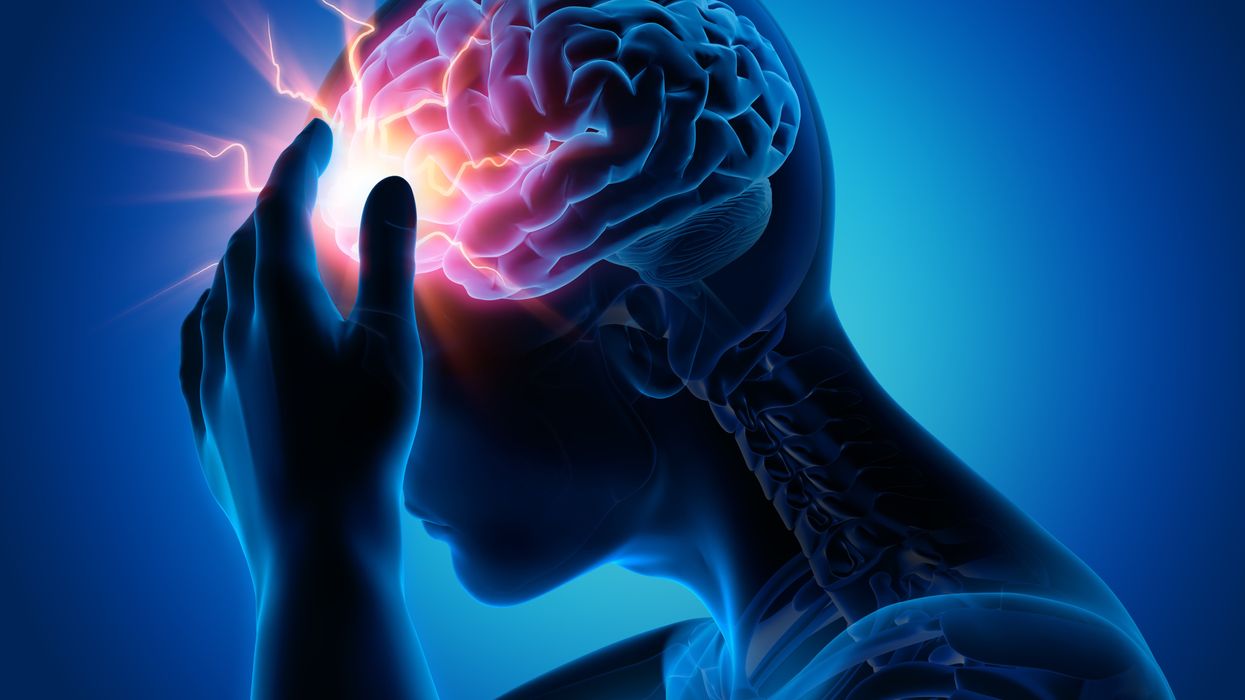
Concussion affects around 42 million people worldwide. While it’s increasingly common in the news because of sports injuries, anything that causes damage to the head, from a fall to a car accident, can result in a concussion. The sudden blow or jolt can disrupt the normal way the brain works. In the immediate aftermath, people may suffer from headaches, lose consciousness and experience dizziness, confusion and vomiting. Some recover but others have side effects that can last for years, particularly affecting memory and concentration.
There is no simple standard-of-care test to confirm a concussion or rule it out. Neither do they appear on MRI and CT scans. Instead, medical professionals use more indirect approaches that test symptoms of concussions, such as assessments of patients’ learning and memory skills, ability to concentrate and problem solving. They also look at balance and coordination. Most tests are in the form of questionnaires or symptom checklists. Consequently, they have limitations, can be biased and may miss a concussion or produce a false positive. Some people suspected of having a concussion may ordinarily have difficulties with literary and problem-solving tests because of language challenges or education levels.
Another problem with current tests is that patients, particularly soldiers who want to return to combat and athletes who would like to keep competing, could try and hide their symptoms to avoid being diagnosed with a brain injury. Trauma physicians who work with concussion patients have the need for a tool that is more objective and consistent.
“This type of assessment doesn’t rely on the patient's education level, willingness to follow instructions or cooperation. You can’t game this.” -- Uzma Samadani, founder of Oculogica
“The importance of having an objective measurement tool for the diagnosis of concussion is of great importance,” says Douglas Powell, associate professor of biomechanics at the University of Memphis, with research interests in sports injury and concussion. “While there are a number of promising systems or metrics, we have yet to develop a system that is portable, accessible and objective for use on the sideline and in the clinic. The EyeBOX may be able to address these issues, though time will be the ultimate test of performance.”
The EyeBOX as a window inside the brain
Using eye movements to diagnose a concussion has emerged as a promising technique since around 2010. Oculogica combined eye movements with AI to develop the EyeBOX to develop an unbiased objective diagnostic tool.
“What’s so great about this type of assessment is it doesn’t rely on the patient's education level, willingness to follow instructions or cooperation,” says Uzma Samadani, a neurosurgeon and brain injury researcher at the University of Minnesota, who founded Oculogica. “You can’t game this. It assesses functions that are prompted by your brain.”
In 2010, Samadani was working on a clinical trial to improve the outcome of brain injuries. The team needed some way to measure if seriously brain injured patients were improving. One thing patients could do was watch TV. So Samadani designed and patented an AI-based algorithm that tracks the relationship between eye movement and concussion.
The EyeBOX test requires patients to watch movie or music clips for 220 seconds. An eye tracking camera records subconscious eye movements, tracking eye positions 500 times per seconds as patients watch the video. It collects over 100,000 data points. The device then uses AI to assess whether there’s any disruptions from the normal way the eyes move.
Cranial nerves are responsible for transmitting information between the brain and the body. Many are involved in eye movement. Pressure caused by a concussion can affect how these nerves work. So tracking how the eyes move can indicate if there’s anything wrong with the cranial nerves and where the problem lies.
If someone is healthy, their eyes should be able to focus on an object, follow movement and both eyes should be coordinated with each other. The EyeBox can detect abnormalities. For example, if a patient’s eyes are coordinated but they are not moving as they should, that indicates issues in the central brain stem, whilst only one eye moving abnormally suggests that a particular nerve section is affected.
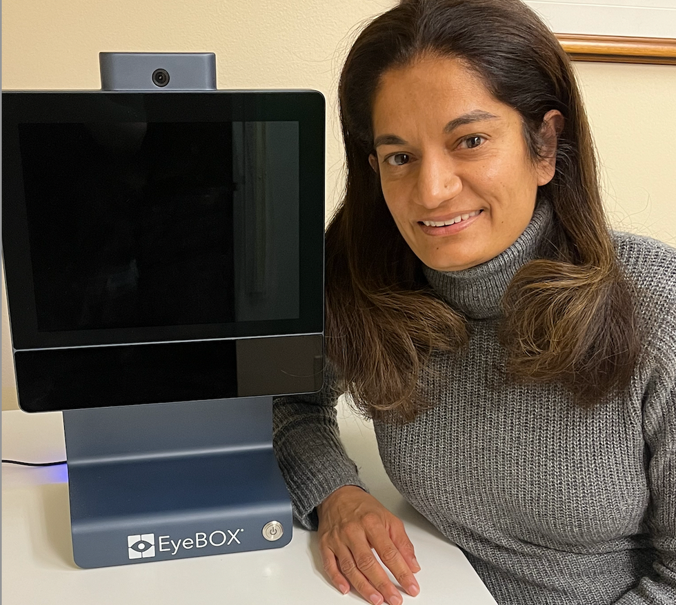
Uzma Samadani with the EyeBOX device
Courtesy Oculogica
“The EyeBOX is a monitor for cranial nerves,” says Samadani. “Essentially it’s a form of digital neurological exam. “Several other eye-tracking techniques already exist, but they rely on subjective self-reported symptoms. Many also require a baseline, a measure of how patients reacted when they were healthy, which often isn’t available.
VOMS (Vestibular Ocular Motor Screen) is one of the most accurate diagnostic tests used in clinics in combination with other tests, but it is subjective. It involves a therapist getting patients to move their head or eyes as they focus or follow a particular object. Patients then report their symptoms.
The King-Devick test measures how fast patients can read numbers and compares it to a baseline. Since it is mainly used for athletes, the initial test is completed before the season starts. But participants can manipulate it. It also cannot be used in emergency rooms because the majority of patients wouldn’t have prior baseline tests.
Unlike these tests, EyeBOX doesn’t use a baseline and is objective because it doesn’t rely on patients’ answers. “It shows great promise,” says Thomas Wilcockson, a senior lecturer of psychology in Loughborough University, who is an expert in using eye tracking techniques in neurological disorders. “Baseline testing of eye movements is not always possible. Alternative measures of concussion currently in development, including work with VR headsets, seem to currently require it. Therefore the EyeBOX may have an advantage.”
A technology that’s still evolving
In their last clinical trial, Oculogica used the EyeBOX to test 46 patients who had concussion and 236 patients who did not. The sensitivity of the EyeBOX, or the probability of it correctly identifying the patient’s concussion, was 80.4 percent. Meanwhile, the test accurately ruled out a concussion in 66.1 percent of cases. This is known as its specificity score.
While the team is working on improving the numbers, experts who treat concussion patients find the device promising. “I strongly support their use of eye tracking for diagnostic decision making,” says Douglas Powell. “But for diagnostic tests, we would prefer at least one of the sensitivity or specificity values to be greater than 90 percent. Powell compares EyeBOX with the Buffalo Concussion Treadmill Test, which has sensitivity and specificity values of 73 and 78 percent, respectively. The VOMS also has shown greater accuracy than the EyeBOX, at least for now. Still, EyeBOX is competitive with the best diagnostic testing available for concussion and Powell hopes that its detection prowess will improve. “I anticipate that the algorithms being used by Oculogica will be under continuous revision and expect the results will improve within the next several years.”
“The color of your skin can have a huge impact in how quickly you are triaged and managed for brain injury. People of color have significantly worse outcomes after traumatic brain injury than people who are white.” -- Uzma Samadani, founder of Oculogica
Powell thinks the EyeBOX could be an important complement to other concussion assessments.
“The Oculogica product is a viable diagnostic tool that supports clinical decision making. However, concussion is an injury that can present with a wide array of symptoms, and the use of technology such as the Oculogica should always be a supplement to patient interaction.”
Ioannis Mavroudis, a consultant neurologist at Leeds Teaching Hospital, agrees that the EyeBOX has promise, but cautions that concussions are too complex to rely on the device alone. For example, not all concussions affect how eyes move. “I believe that it can definitely help, however not all concussions show changes in eye movements. I believe that if this could be combined with a cognitive assessment the results would be impressive.”
The Oculogica team submitted their clinical data for FDA approval and received it in 2018. Now, they’re working to bring the test to the commercial market and using the device clinically to help diagnose concussions for clients. They also want to look at other areas of brain health in the next few years. Samadani believes that the EyeBOX could possibly be used to detect diseases like multiple sclerosis or other neurological conditions. “It’s a completely new way of figuring out what someone’s neurological exam is and we’re only beginning to realize the potential,” says Samadani.
One of Samadani’s biggest aspirations is to help reduce inequalities in healthcare because of skin color and other factors like money or language barriers. From that perspective, the EyeBOX’s greatest potential could be in emergency rooms. It can help diagnose concussions in addition to the questionnaires, assessments and symptom checklists, currently used in the emergency departments. Unlike these more subjective tests, EyeBOX can produce an objective analysis of brain injury through AI when patients are admitted and assessed, unrelated to their socioeconomic status, education, or language abilities. Studies suggest that there are racial disparities in how patients with brain injuries are treated, such as how quickly they're assessed and get a treatment plan.
“The color of your skin can have a huge impact in how quickly you are triaged and managed for brain injury,” says Samadani. “As a result of that, people of color have significantly worse outcomes after traumatic brain injury than people who are white. The EyeBOX has the potential to reduce inequalities,” she explains.
“If you had a digital neurological tool that you could screen and triage patients on admission to the emergency department you would potentially be able to make sure that everybody got the same standard of care,” says Samadani. “My goal is to change the way brain injury is diagnosed and defined.”