Scientists make progress with growing organs for transplants

Researchers from the University of Cambridge have laid the foundations for growing synthetic embryos that could develop a beating heart, gut and brain.
Story by Big Think
For over a century, scientists have dreamed of growing human organs sans humans. This technology could put an end to the scarcity of organs for transplants. But that’s just the tip of the iceberg. The capability to grow fully functional organs would revolutionize research. For example, scientists could observe mysterious biological processes, such as how human cells and organs develop a disease and respond (or fail to respond) to medication without involving human subjects.
Recently, a team of researchers from the University of Cambridge has laid the foundations not just for growing functional organs but functional synthetic embryos capable of developing a beating heart, gut, and brain. Their report was published in Nature.
The organoid revolution
In 1981, scientists discovered how to keep stem cells alive. This was a significant breakthrough, as stem cells have notoriously rigorous demands. Nevertheless, stem cells remained a relatively niche research area, mainly because scientists didn’t know how to convince the cells to turn into other cells.
Then, in 1987, scientists embedded isolated stem cells in a gelatinous protein mixture called Matrigel, which simulated the three-dimensional environment of animal tissue. The cells thrived, but they also did something remarkable: they created breast tissue capable of producing milk proteins. This was the first organoid — a clump of cells that behave and function like a real organ. The organoid revolution had begun, and it all started with a boob in Jello.
For the next 20 years, it was rare to find a scientist who identified as an “organoid researcher,” but there were many “stem cell researchers” who wanted to figure out how to turn stem cells into other cells. Eventually, they discovered the signals (called growth factors) that stem cells require to differentiate into other types of cells.
For a human embryo (and its organs) to develop successfully, there needs to be a “dialogue” between these three types of stem cells.
By the end of the 2000s, researchers began combining stem cells, Matrigel, and the newly characterized growth factors to create dozens of organoids, from liver organoids capable of producing the bile salts necessary for digesting fat to brain organoids with components that resemble eyes, the spinal cord, and arguably, the beginnings of sentience.
Synthetic embryos
Organoids possess an intrinsic flaw: they are organ-like. They share some characteristics with real organs, making them powerful tools for research. However, no one has found a way to create an organoid with all the characteristics and functions of a real organ. But Magdalena Żernicka-Goetz, a developmental biologist, might have set the foundation for that discovery.
Żernicka-Goetz hypothesized that organoids fail to develop into fully functional organs because organs develop as a collective. Organoid research often uses embryonic stem cells, which are the cells from which the developing organism is created. However, there are two other types of stem cells in an early embryo: stem cells that become the placenta and those that become the yolk sac (where the embryo grows and gets its nutrients in early development). For a human embryo (and its organs) to develop successfully, there needs to be a “dialogue” between these three types of stem cells. In other words, Żernicka-Goetz suspected the best way to grow a functional organoid was to produce a synthetic embryoid.
As described in the aforementioned Nature paper, Żernicka-Goetz and her team mimicked the embryonic environment by mixing these three types of stem cells from mice. Amazingly, the stem cells self-organized into structures and progressed through the successive developmental stages until they had beating hearts and the foundations of the brain.
“Our mouse embryo model not only develops a brain, but also a beating heart [and] all the components that go on to make up the body,” said Żernicka-Goetz. “It’s just unbelievable that we’ve got this far. This has been the dream of our community for years and major focus of our work for a decade and finally we’ve done it.”
If the methods developed by Żernicka-Goetz’s team are successful with human stem cells, scientists someday could use them to guide the development of synthetic organs for patients awaiting transplants. It also opens the door to studying how embryos develop during pregnancy.
This article originally appeared on Big Think, home of the brightest minds and biggest ideas of all time.

Small changes in how a person talks could reveal Alzheimer’s earlier
In an initial study, Canary analyzed speech recordings with AI and identified early stage Alzheimer’s with 96 percent accuracy.
Dave Arnold retired in his 60s and began spending time volunteering in local schools. But then he started misplacing items, forgetting appointments and losing his sense of direction. Eventually he was diagnosed with early stage Alzheimer’s.
“Hearing the diagnosis made me very emotional and tearful,” he said. “I immediately thought of all my mom had experienced.” His mother suffered with the condition for years before passing away. Over the last year, Arnold has worked for the Alzheimer’s Association as one of its early stage advisors, sharing his insights to help others in the initial stages of the disease.
Arnold was diagnosed sooner than many others. It's important to find out early, when interventions can make the most difference. One promising avenue is looking at how people talk. Research has shown that Alzheimer’s affects a part of the brain that controls speech, resulting in small changes before people show other signs of the disease.
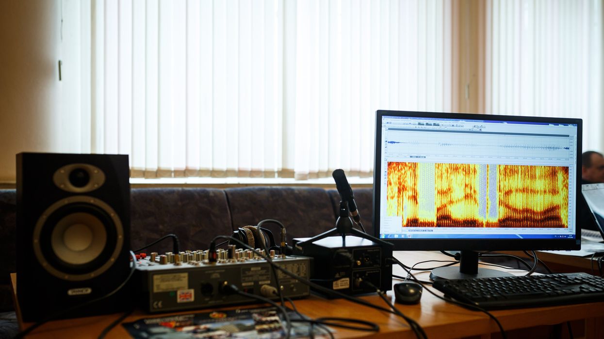
Now, Canary Speech, a company based in Utah, is using AI to examine elements like the pitch of a person’s voice and their pauses. In an initial study, Canary analyzed speech recordings with AI and identified early stage Alzheimer’s with 96 percent accuracy.
Developing the AI model
Canary Speech’s CEO, Henry O’Connell, met cofounder Jeff Adams about 40 years before they started the company. Back when they first crossed paths, they were both living in Bethesda, Maryland; O’Connell was a research fellow at the National Institutes of Health studying rare neurological diseases, while Adams was working to decode spy messages. Later on, Adams would specialize in building mathematical models to analyze speech and sound as a team leader in developing Amazon's Alexa.
It wasn't until 2015 that they decided to make use of the fit between their backgrounds. ““We established Canary Speech in 2017 to build a product that could be used in multiple languages in clinical environments,” O'Connell says.
The need is growing. About 55 million people worldwide currently live with Alzheimer’s, a number that is expected to double by 2050. Some scientists think the disease results from a buildup of plaque in the brain. It causes mild memory loss at first and, over time, this issue get worse while other symptoms, such as disorientation and hallucinations, can develop. Treatment to manage the disease is more effective in the earlier stages, but detection is difficult since mild symptoms are often attributed to the normal aging process.
O’Connell and Adams specialize in the complex ways that Alzheimer’s effects how people speak. Using AI, their mathematical model analyzes 15 million data points every minute, focusing on certain features of speech such as pitch, pauses and elongation of words. It also pays attention to how the vibrations of vocal cords change in different stages of the disease.
To create their model, the team used a type of machine learning called deep neural nets, which looks at multiple layers of data - in this case, the multiple features of a person’s speech patterns.
“Deep neural nets allow us to look at much, much larger data sets built out of millions of elements,” O’Connell explained. “Through machine learning and AI, we’ve identified features that are very sensitive to an Alzheimer’s patient versus [people without the disease] and also very sensitive to mild cognitive impairment, early stage and moderate Alzheimer's.” Based on their learnings, Canary is able to classify the disease stage very quickly, O’Connell said.
“When we’re listening to sublanguage elements, we’re really analyzing the direct result of changes in the brain in the physical body,” O’Connell said. “The brain controls your vocal cords: how fast they vibrate, the expansion of them, the contraction.” These factors, along with where people put their tongues when talking, function subconsciously and result in subtle changes in the sounds of speech.
Further testing is needed
In an initial trial, Canary analyzed speech recordings from phone calls to a large U.S. health insurer. They looked at the audio recordings of 651 policyholders who had early stage Alzheimer’s and 1018 who did not have the condition, aiming for a representative sample of age, gender and race. They used this data to create their first diagnostic model and found that it was 96 percent accurate in identifying Alzheimer’s.
Christian Herff, an assistant professor of neuroscience at Maastricht University in the Netherlands, praised this approach while adding that further testing is needed to assess its effectiveness.
“I think the general idea of identifying increased risk for cognitive impairment based on speech characteristics is very feasible, particularly when change in a user’s voice is monitored, for example, by recording speech every year,” Herff said. He noted that this can only be a first indication, not a full diagnosis. The accuracy still needs to be validated in studies that follows individuals over a period of time, he said.
Toby Walsh, a professor of artificial intelligence at the University of New South Wales, also thinks Canary’s tool has potential but highlights that Canary could diagnose some people who don’t really have the disease. “This is an interesting and promising application of AI,” he said, “but these tools need to be used carefully. Imagine the anxiety of being misdiagnosed with Alzheimer’s.”
As with many other AI tools, privacy and bias are additional issues to monitor closely, Walsh said.
Other languages
A related issue is that not everyone is fluent in English. Mahnaz Arvaneh, a senior lecturer in automatic control and systems engineering at the University of Sheffield, said this could be a blind spot.
“The system may not be very accurate for those who have English as their second language as their speaking patterns would be different, and any issue might be because of language deficiency rather than cognitive issues,” Arvaneh said.
The team is expanding to multiple languages starting with Japanese and Spanish. The elements of the model that make up the algorithm are very similar, but they need to be validated and retrained in a different language, which will require access to more data.
Recently, Canary analyzed the phone calls of 233 Japanese patients who had mild cognitive impairment and 704 healthy people. Using an English model they were able to identify the Japanese patients who had mild cognitive impairment with 78 percent accuracy. They also developed a model in Japanese that was 45 percent accurate, and they’re continuing to train it with more data.
The future
Canary is using their model to look at other diseases like Huntington’s and Parkinson’s. They’re also collaborating with pharmaceuticals to validate potential therapies for Alzheimer’s. By looking at speech patterns over time, Canary can get an indication of how well these drugs are working.

Dave Arnold and his wife dance at his nephew’s wedding in Rochester, New York, ten years ago, before his Alzheimer's diagnosis.
Dave Arnold
Ultimately, they want to integrate their tool into everyday life. “We want it to be used in a smartphone, or a teleconference call so that individuals could be examined in their home,” O’Connell said. “We could follow them over time and work with clinical teams and hospitals to improve the evaluation of patients and contribute towards an accurate diagnosis.”
Arnold, the patient with early stage Alzheimer’s, sees great promise. “The process of getting a diagnosis is already filled with so much anxiety,” he said. “Anything that can be done to make it easier and less stressful would be a good thing, as long as it’s proven accurate.”
Gene therapy helps restore teen’s vision for first time
Doctors used new eye drops to treat a rare genetic disorder.
Story by Freethink
For the first time, a topical gene therapy — designed to heal the wounds of people with “butterfly skin disease” — has been used to restore a person’s vision, suggesting a new way to treat genetic disorders of the eye.
The challenge: Up to 125,000 people worldwide are living with dystrophic epidermolysis bullosa (DEB), an incurable genetic disorder that prevents the body from making collagen 7, a protein that helps strengthen the skin and other connective tissues.Without collagen 7, the skin is incredibly fragile — the slightest friction can lead to the formation of blisters and scarring, most often in the hands and feet, but in severe cases, also the eyes, mouth, and throat.
This has earned DEB the nickname of “butterfly skin disease,” as people with it are said to have skin as delicate as a butterfly’s wings.
The gene therapy: In May 2023, the FDA approved Vyjuvek, the first gene therapy to treat DEB.
Vyjuvek uses an inactivated herpes simplex virus to deliver working copies of the gene for collagen 7 to the body’s cells. In small trials, 65 percent of DEB-caused wounds sprinkled with it healed completely, compared to just 26 percent of wounds treated with a placebo.
“It was like looking through thick fog.” -- Antonio Vento Carvajal.
The patient: Antonio Vento Carvajal, a 14 year old living in Florida, was one of the trial participants to benefit from Vyjuvek, which was developed by Pittsburgh-based pharmaceutical company Krystal Biotech.
While the topical gene therapy could help his skin, though, it couldn’t do anything to address the severe vision loss Antonio experienced due to his DEB. He’d undergone multiple surgeries to have scar tissue removed from his eyes, but due to his condition, the blisters keep coming back.
“It was like looking through thick fog,” said Antonio, noting how his impaired vision made it hard for him to play his favorite video games. “I had to stand up from my chair, walk over, and get closer to the screen to be able to see.”
The idea: Encouraged by how Antonio’s skin wounds were responding to the gene therapy, Alfonso Sabater, his doctor at the Bascom Palmer Eye Institute, reached out to Krystal Biotech to see if they thought an alternative formula could potentially help treat his patient’s eyes.
The company was eager to help, according to Sabater, and after about two years of safety and efficacy testing, he had permission, under the FDA’s compassionate use protocol, to treat Antonio’s eyes with a version of the topical gene therapy delivered as eye drops.
The results: In August 2022, Sabater once again removed scar tissue from Antonio’s right eye, but this time, he followed up the surgery by immediately applying eye drops containing the gene therapy.
“I would send this message to other families in similar situations, whether it’s DEB or another condition that can benefit from genetic therapy. Don’t be afraid.” -- Yunielkys “Yuni” Carvajal.
The vision in Antonio’s eye steadily improved. By about eight months after the treatment, it was just slightly below average (20/25) and stayed that way. In March 2023, Sabater performed the same procedure on his young patient’s other eye, and the vision in it has also steadily improved.
“I’ve seen the transformation in Antonio’s life,” said Sabater. “He’s always been a happy kid. Now he’s very happy. He can function pretty much normally. He can read, he can study, he can play video games.”
Looking ahead: The topical gene therapy isn’t a permanent fix — it doesn’t alter Antonio’s own genes, so he has to have the eye drops reapplied every month. Still, that’s far less invasive than having to undergo repeated surgeries.
Sabater is now working with Krystal Biotech to launch trials of the eye drops in other patients, and not just those with DEB. By changing the gene delivered by the therapy, he believes it could be used to treat other eye disorders that are far more common — Fuchs’ dystrophy, for example, affects the vision of an estimated 300 million people over the age of 30.
Antonio’s mother, Yunielkys “Yuni” Carvajal, meanwhile, has said that having her son be the first to receive the eye drops was “very scary,” but she’s hopeful others will take a chance on new gene therapies if given the opportunity.
“I would send this message to other families in similar situations, whether it’s DEB or another condition that can benefit from genetic therapy,” she said. “Don’t be afraid.”
This article originally appeared on Freethink, home of the brightest minds and biggest ideas of all time.


