The Scientist Behind the Pap Smear Saved Countless Women from Cervical Cancer

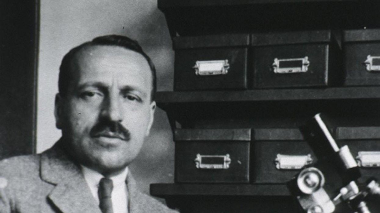
George Papanicolaou (1883-1962), Greek-born American physician developed a simple cytological test for cervical cancer in 1928.
For decades, women around the world have made the annual pilgrimage to their doctor for the dreaded but potentially life-saving Papanicolaou test, a gynecological exam to screen for cervical cancer named for Georgios Papanicolaou, the Greek immigrant who developed it.
The Pap smear, as it is commonly known, is credited for reducing cervical cancer mortality by 70% since the 1960s; the American Cancer Society (ACS) still ranks the Pap as the most successful screening test for preventing serious malignancies. Nonetheless, the agency, as well as other medical panels, including the US Preventive Services Task Force and the American College of Obstetrics and Gynecology are making a strong push to replace the Pap with the more sensitive high-risk HPV screening test for the human papillomavirus virus, which causes nearly all cases of cervical cancer.
So, how was the Pap developed and how did it become the gold standard of cervical cancer detection for more than 60 years?
Born on May 13, 1883, on the island of Euboea, Greece, Georgios Papanicolaou attended the University of Athens where he majored in music and the humanities before earning his medical degree in 1904 and PhD from the University of Munich six years later. In Europe, Papanicolaou was an assistant military surgeon during the Balkan War, a psychologist for an expedition of the Oceanographic Institute of Monaco and a caregiver for leprosy patients.
When he and his wife, Andromache Mavroyenous (Mary), arrived at Ellis Island on October 19, 1913, the young couple had scarcely more than the $250 minimum required to immigrate, spoke no English and had no job prospects. They worked a series of menial jobs--department store sales clerk, rug salesman, newspaper clerk, restaurant violinist--before Papanicolaou landed a position as an anatomy assistant at Cornell University and Mary was hired as his lab assistant, an arrangement that would last for the next 50 years.
Papanikolaou would later say the discovery "was one of the greatest thrills I ever experienced during my scientific career."
In his early research, Papanikolaou used guinea pigs to prove that gender is determined by the X and Y chromosomes. Using a pediatric nasal speculum, he collected and microscopically examined vaginal secretions of guinea pigs, which revealed distinct cell changes connected to the menstrual cycle. He moved on to study reproductive patterns in humans, beginning with his faithful wife, Mary, who not only endured his almost-daily cervical exams for decades, but also recruited friends as early research participants.
Writing in the medical journal Growth in 1920, the scientist outlined his theory that a microscopic smear of vaginal fluid could detect the presence of cancer cells in the uterus. Papanikolaou would later say the discovery "was one of the greatest thrills I ever experienced during my scientific career."
At this time, cervical cancer was the number one cancer killer of American women but physicians were skeptical of these new findings. They continued to rely on biopsy and curettage to diagnose and treat the disease until Papanicolaou's discovery was published in American Journal of Obstetrics and Gynecology. An inexpensive, easy-to-perform test that could detect cervical cancer, precancerous dysplasia and other cytological diseases was a sea change. Between 1975 and 2001, the cervical cancer rate was cut in half.
Papanicolaou became Emeritus Professor at Cornell University Medical College and received numerous awards, including the Albert Lasker Award for Clinical Medical Research and the Medal of Honor from the American Cancer Society. His image was featured on the Greek currency and the US Post Office issued a commemorative stamp in his honor. But international acclaim didn't lead to a more relaxed schedule. The researcher continued to work seven days a week and refused to take vacations.
After nearly 50 years, Papanicolaou left Cornell to head and develop the Cancer Institute of Miami. He died of a heart attack on February 19, 1962, just three months after his arrival. Mary continued to work in the renamed Papanicolaou Cancer Research Institute until her death 20 years later.
The annual pap smear was originally tied to renewing a birth control prescription. Canada began recommending Pap exams every three years in 1978. The United States followed suit in 2012, noting that it takes many years for cervical cancer to develop. In September 2020, the American Cancer Society recommended delaying the first gynecological pelvic exam until age 25 and replacing the Pap test completely with the more accurate human papillomavirus (HPV) test every five years as the technology becomes more widely available.
Not everyone agrees that it's time to do away with this proven screening method, though. The incidence rate of cervical cancer among Hispanic women is 28% higher than for white women, and Black women are more likely to die of cervical cancer than any other racial or ethnicities.
Whether the Pap is administered every year, every three years or not at all, Papanicolaou will always be known as the medical hero who saved countless women who would otherwise have succumbed to cervical cancer.
The Pandemic Is Ushering in a More Modern—and Ethical—Way of Studying New Drugs and Diseases
Miniature lab-grown models of human organs have major advantages over standard animal testing.
Before the onset of the coronavirus pandemic, Dutch doctoral researcher Joep Beumer had used miniature lab-grown organs to study the human intestine as part of his PhD thesis. When lockdown hit, however, he was forced to delay his plans for graduation. Overwhelmed by a sense of boredom after the closure of his lab at the Hubrecht Institute, in the Netherlands, he began reading literature related to COVID-19.
"By February [2020], there were already reports on coronavirus symptoms in the intestinal tract," Beumer says, adding that this piqued his interest. He wondered if he could use his miniature models – called organoids -- to study how the coronavirus infects the intestines.
But he wasn't the only one to follow this train of thought. In the year since the pandemic began, many researchers have been using organoids to study how the coronavirus infects human cells, and find potential treatments. Beumer's pivot represents a remarkable and fast-emerging paradigm shift in how drugs and diseases will be studied in the coming decades. With future pandemics likely to be more frequent and deadlier, such a shift is necessary to reduce the average clinical development time of 5.9 years for antiviral agents.
Part of that shift means developing models that replicate human biology in the lab. Animal models, which are the current standard in biomedical research, fail to do so—96% of drugs that pass animal testing, for example, fail to make it to market. Injecting potentially toxic drugs into living creatures, before eventually slaughtering them, also raises ethical concerns for some. Organoids, on the other hand, respond to infectious diseases, or potential treatments, in a way that is relevant to humans, in addition to being slaughter-free.

Human intestinal organoids infected with SARS-CoV-2 (white).
Credit: Joep Beumer/Clevers group/Hubrecht Institute
Urgency Sparked Momentum
Though brain organoids were previously used to study the Zika virus during the 2015-16 epidemic, it wasn't until COVID-19 that the field really started to change. "The organoid field has advanced a lot in the last year. The speed at which it happened is crazy," says Shuibing Chen, an associate professor at Weill Cornell Medicine in New York. She adds that many federal and private funding agencies have now seen the benefits of organoids, and are starting to appreciate their potential in the biomedical field.
Last summer, the Organo-Strat (OS) network—a German network that uses human organoid models to study COVID-19's effects—received 3.2 million euros in funding from the German government. "When the pandemic started, we became aware that we didn't have the right models to immediately investigate the effects of the virus," says Andreas Hocke, professor of infectious diseases at the Charité Universitätsmedizin in Berlin, Germany, and coordinator of the OS network. Hocke explained that while the World Health Organization's animal models showed an "overlap of symptoms'' with humans, there was "no clear reflection" of the same disease.
"The network functions as a way of connecting organoid experts with infectious disease experts across Germany," Hocke continues. "Having organoid models on demand means we can understand how a virus infects human cells from the first moment it's isolated." Overall, OS aims to create infrastructure that could be applied to future pandemics. There are 28 sub-projects involved in the network, covering a wide assortment of individual organoids.
Cost, however, remains an obstacle to scaling up, says Chen. She says there is also a limit to what we can learn from organoids, given that they only represent a single organ. "We can add drugs to organoids to see how the cells respond, but these tests don't tell us anything about drug metabolism, for example," she explains.
A Related "Leaps" in Progress
One way to solve this issue is to use an organ-on-a-chip system. These are miniature chips containing a variety of human cells, as well as small channels along which functions like blood or air flow can be recreated. This allows scientists to perform more complex experiments, like studying drug metabolism, while producing results that are relevant to humans.

An organ-on-a-chip system.
Credit: Fraunhofer IGB
Such systems are also able to elicit an immune response. The FDA has even entered into an agreement with Wyss Institute spinoff Emulate to use their lung-on-a-chip system to test COVID-19 vaccines. Representing multiple organs in one system is also possible. Berlin-based TissUse are aiming to make a so-called 'human on a chip' system commercially available. But TissUse senior scientist Ilka Maschmeyer warns that there is a limit to how far the technology can go. "The system will not think or feel, so it wouldn't be possible to test for illnesses affecting these abilities," she says.
Some challenges also remain in the usability of organs-on-a-chip. "Specialized training is required to use them as they are so complex," says Peter Loskill, assistant professor and head of the organ-on-a-chip group at the University of Tübingen, Germany. Hocke agrees with this. "Cell culture scientists would easily understand how to use organoids in a lab, but when using a chip, you need additional biotechnology knowledge," he says.
One major advantage of both technologies is the possibility of personalized medicine: Cells can be taken from a patient and put onto a chip, for example, to test their individual response to a treatment. Loskill also says there are other uses outside of the biomedical field, such as cosmetic and chemical testing.
"Although these technologies offer a lot of possibilities, they need time to develop," Loskill continues. He stresses, however, that it's not just the technology that needs to change. "There's a lot of conservative thinking in biomedical research that says this is how we've always done things. To really study human biology means approaching research questions in a completely new way."
Even so, he thinks that the pandemic marked a shift in people's thinking—no one cared how the results were found, as long as it was done quickly. But Loskill adds that it's important to balance promise, potential, and expectations when it comes to these new models. "Maybe in 15 years' time we will have a limited number of animal models in comparison to now, but the timescale depends on many factors," he says.
Beumer, now a post-doc, was eventually allowed to return to the lab to develop his coronavirus model, and found working on it to be an eye-opening experience. He saw first-hand how his research could have an impact on something that was affecting the entire human race, as well as the pressure that comes with studying potential treatments. Though he doesn't see a future for himself in infectious diseases, he hopes to stick with organoids. "I've now gotten really excited about the prospect of using organoids for drug discovery," he says.
The coronavirus pandemic has slowed society down in many respects, but it has flung biomedical research into the future—from mRNA vaccines to healthcare models based on human biology. It may be difficult to fully eradicate animal models, but over the coming years, organoids and organs-on-a-chip may become the standard for the sake of efficacy -- and ethics.
Jack McGovan is a freelance science writer based in Berlin. His main interests center around sustainability, food, and the multitude of ways in which the human world intersects with animal life. Find him on Twitter @jack_mcgovan."
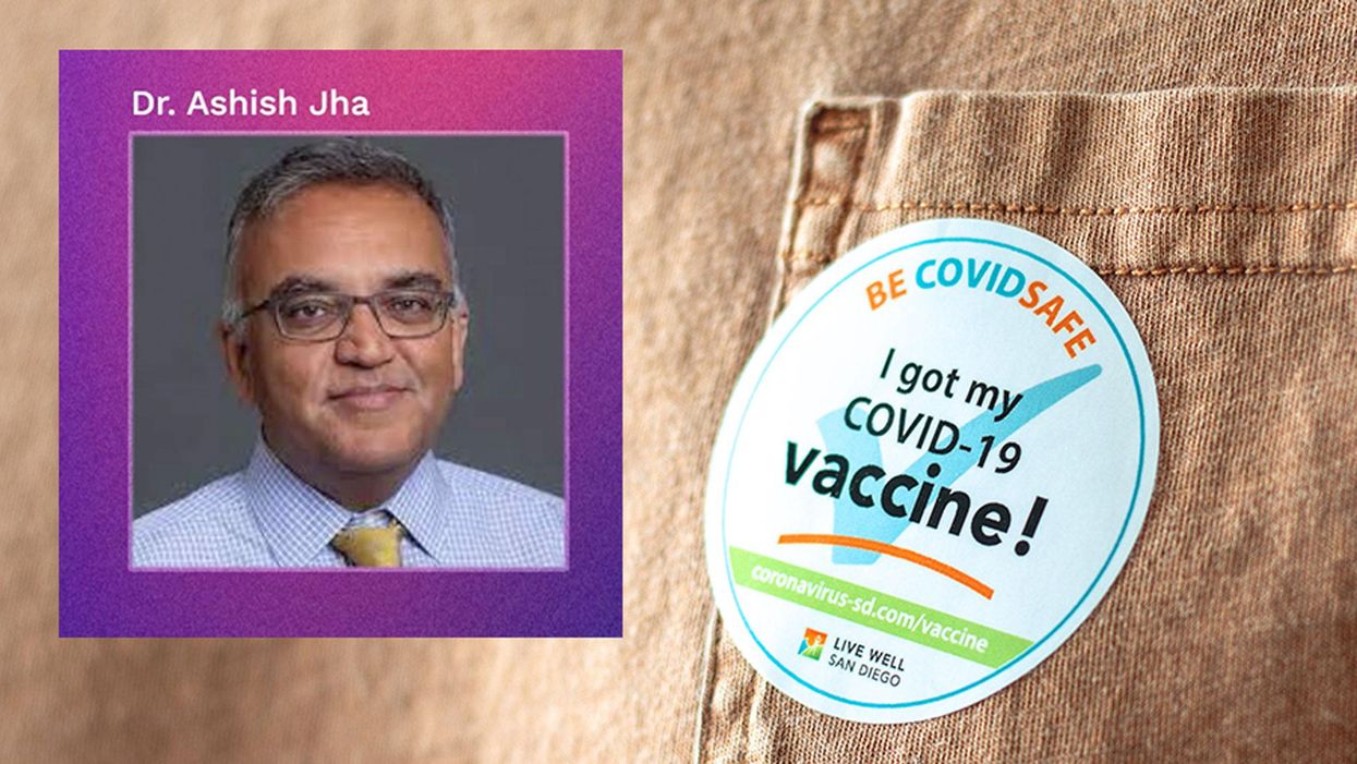
New Podcast: Why Dr. Ashish Jha Expects a Good Summer
Dr. Jha discusses Covid vaccine passports, how supply and demand of the vaccines is about to shift, the AstraZeneca situation, what's new with kids, herd immunity, and more.
Making Sense of Science features interviews with leading medical and scientific experts about the latest developments and the big ethical and societal questions they raise. This monthly podcast is hosted by journalist Kira Peikoff, founding editor of the award-winning science outlet Leaps.org.
Hear the 30-second trailer:
Listen to the whole episode: "Why Dr. Ashish Jha Expects a Good Summer"
Dr. Ashish Jha, dean of public health at Brown University, discusses the latest developments around the Covid-19 vaccines, including supply and demand, herd immunity, kids, vaccine passports, and why he expects the summer to look very good.
Kira Peikoff was the editor-in-chief of Leaps.org from 2017 to 2021. As a journalist, her work has appeared in The New York Times, Newsweek, Nautilus, Popular Mechanics, The New York Academy of Sciences, and other outlets. She is also the author of four suspense novels that explore controversial issues arising from scientific innovation: Living Proof, No Time to Die, Die Again Tomorrow, and Mother Knows Best. Peikoff holds a B.A. in Journalism from New York University and an M.S. in Bioethics from Columbia University. She lives in New Jersey with her husband and two young sons. Follow her on Twitter @KiraPeikoff.

