Researchers Are Testing a New Stem Cell Therapy in the Hopes of Saving Millions from Blindness

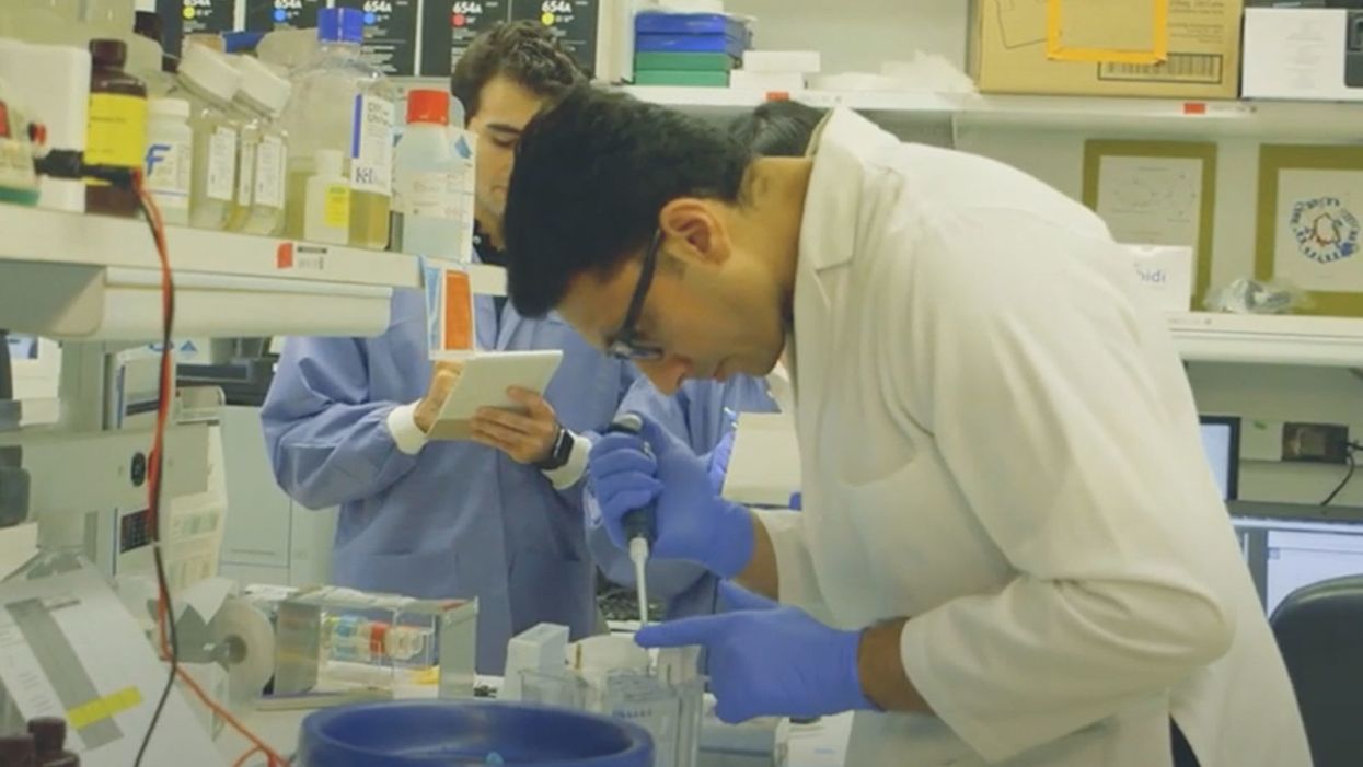
NIH researchers in Kapil Bharti's lab work toward the development of induced pluripotent stem cells to treat dry age-related macular degeneration.
Of all the infirmities of old age, failing sight is among the cruelest. It can mean the end not only of independence, but of a whole spectrum of joys—from gazing at a sunset or a grandchild's face to reading a novel or watching TV.
The Phase 1 trial will likely run through 2022, followed by a larger Phase 2 trial that could last another two or three years.
The leading cause of vision loss in people over 55 is age-related macular degeneration, or AMD, which afflicts an estimated 11 million Americans. As photoreceptors in the macula (the central part of the retina) die off, patients experience increasingly severe blurring, dimming, distortions, and blank spots in one or both eyes.
The disorder comes in two varieties, "wet" and "dry," both driven by a complex interaction of genetic, environmental, and lifestyle factors. It begins when deposits of cellular debris accumulate beneath the retinal pigment epithelium (RPE)—a layer of cells that nourish and remove waste products from the photoreceptors above them. In wet AMD, this process triggers the growth of abnormal, leaky blood vessels that damage the photoreceptors. In dry AMD, which accounts for 80 to 90 percent of cases, RPE cells atrophy, causing photoreceptors to wither away. Wet AMD can be controlled in about a quarter of patients, usually by injections of medication into the eye. For dry AMD, no effective remedy exists.
Stem Cells: Promise and Perils
Over the past decade, stem cell therapy has been widely touted as a potential treatment for AMD. The idea is to augment a patient's ailing RPE cells with healthy ones grown in the lab. A few small clinical trials have shown promising results. In a study published in 2018, for example, a University of Southern California team cultivated RPE tissue from embryonic stem cells on a plastic matrix and transplanted it into the retinas of four patients with advanced dry AMD. Because the trial was designed to test safety rather than efficacy, lead researcher Amir Kashani told a reporter, "we didn't expect that replacing RPE cells would return a significant amount of vision." Yet acuity improved substantially in one recipient, and the others regained their lost ability to focus on an object.
Therapies based on embryonic stem cells, however, have two serious drawbacks: Using fetal cell lines raises ethical issues, and such treatments require the patient to take immunosuppressant drugs (which can cause health problems of their own) to prevent rejection. That's why some experts favor a different approach—one based on induced pluripotent stem cells (iPSCs). Such cells, first produced in 2006, are made by returning adult cells to an undifferentiated state, and then using chemicals to reprogram them as desired. Treatments grown from a patient's own tissues could sidestep both hurdles associated with embryonic cells.
At least hypothetically. Today, the only stem cell therapies approved by the U.S. Food and Drug Administration (FDA) are umbilical cord-derived products for various blood and immune disorders. Although scientists are probing the use of embryonic stem cells or iPSCs for conditions ranging from diabetes to Parkinson's disease, such applications remain experimental—or fraudulent, as a growing number of patients treated at unlicensed "stem cell clinics" have painfully learned. (Some have gone blind after receiving bogus AMD therapies at those facilities.)
Last December, researchers at the National Eye Institute in Bethesda, Maryland, began enrolling patients with dry AMD in the country's first clinical trial using tissue grown from the patients' own stem cells. Led by biologist Kapil Bharti, the team intends to implant custom-made RPE cells in 12 recipients. If the effort pans out, it could someday save the sight of countless oldsters.
That, however, is what's technically referred to as a very big "if."
The First Steps
Bharti's trial is not the first in the world to use patient-derived iPSCs to treat age-related macular degeneration. In 2013, Japanese researchers implanted such cells into the eyes of a 77-year-old woman with wet AMD; after a year, her vision had stabilized, and she no longer needed injections to keep abnormal blood vessels from forming. A second patient was scheduled for surgery—but the procedure was canceled after the lab-grown RPE cells showed signs of worrisome mutations. That incident illustrates one potential problem with using stem cells: Under some circumstances, the cells or the tissue they form could turn cancerous.
"The knowledge and expertise we're gaining can be applied to many other iPSC-based therapies."
Bharti and his colleagues have gone to great lengths to avoid such outcomes. "Our process is significantly different," he told me in a phone interview. His team begins with patients' blood stem cells, which appear to be more genomically stable than the skin cells that the Japanese group used. After converting the blood cells to RPE stem cells, his team cultures them in a single layer on a biodegradable scaffold, which helps them grow in an orderly manner. "We think this material gives us a big advantage," Bharti says. The team uses a machine-learning algorithm to identify optimal cell structure and ensure quality control.
It takes about six months for a patch of iPSCs to become viable RPE cells. When they're ready, a surgeon uses a specially-designed tool to insert the tiny structure into the retina. Within days, the scaffold melts away, enabling the transplanted RPE cells to integrate fully into their new environment. Bharti's team initially tested their method on rats and pigs with eye damage mimicking AMD. The study, published in January 2019 in Science Translational Medicine, found that at ten weeks, the implanted RPE cells continued to function normally and protected neighboring photoreceptors from further deterioration. No trace of mutagenesis appeared.
Encouraged by these results, Bharti began recruiting human subjects. The Phase 1 trial will likely run through 2022, followed by a larger Phase 2 trial that could last another two or three years. FDA approval would require an even larger Phase 3 trial, with a decision expected sometime between 2025 and 2028—that is, if nothing untoward happens before then. One unknown (among many) is whether implanted cells can thrive indefinitely under the biochemically hostile conditions of an eye with AMD.
"Most people don't have a sense of just how long it takes to get something like this to work, and how many failures—even disasters—there are along the way," says Marco Zarbin, professor and chair of Ophthalmology and visual science at Rutgers New Jersey Medical School and co-editor of the book Cell-Based Therapy for Degenerative Retinal Diseases. "The first kidney transplant was done in 1933. But the first successful kidney transplant was in 1954. That gives you a sense of the time frame. We're really taking the very first steps in this direction."
Looking Ahead
Even if Bharti's method proves safe and effective, there's the question of its practicality. "My sense is that using induced pluripotent stem cells to treat the patient from whom they're derived is a very expensive undertaking," Zarbin observes. "So you'd have to have a very dramatic clinical benefit to justify that cost."
Bharti concedes that the price of iPSC therapy is likely to be high, given that each "dose" is formulated for a single individual, requires months to manufacture, and must be administered via microsurgery. Still, he expects economies of scale and production to emerge with time. "We're working on automating several steps of the process," he explains. "When that kicks in, a technician will be able to make products for 10 or 20 people at once, so the cost will drop proportionately."
Meanwhile, other researchers are pressing ahead with therapies for AMD using embryonic stem cells, which could be mass-produced to treat any patient who needs them. But should that approach eventually win FDA approval, Bharti believes there will still be room for a technique that requires neither fetal cell lines nor immunosuppression.
And not only for eye ailments. "The knowledge and expertise we're gaining can be applied to many other iPSC-based therapies," says the scientist, who is currently consulting with several companies that are developing such treatments. "I'm hopeful that we can leverage these approaches for a wide range of applications, whether it's for vision or across the body."
NEI launches iPS cell therapy trial for dry AMD
The Cellular Secrets of “Young Blood” Are Starting to Be Unlocked
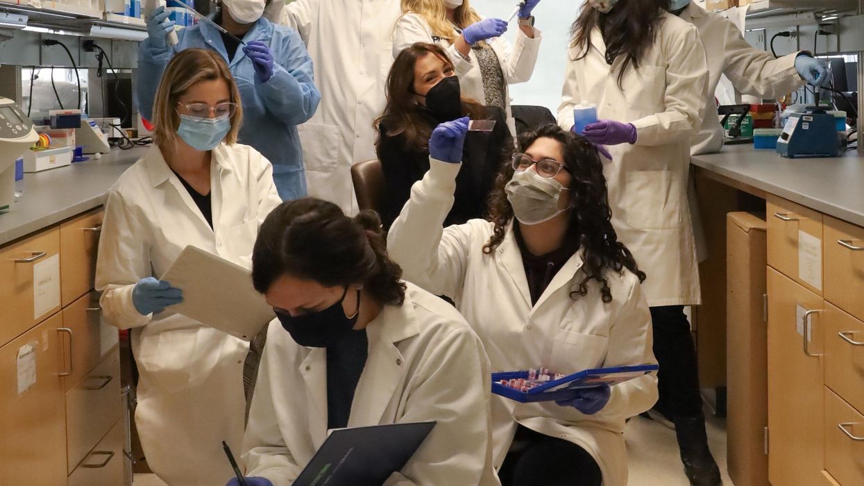
Fabrisia Ambrosio (center) surrounded by her lab staff.
The quest for an elixir to restore youthful health and vigor is common to most cultures and has prompted much scientific research. About a decade ago, Stanford scientists stitched together the blood circulatory systems of old and young mice in a practice called parabiosis. It seemed to rejuvenate the aged animals and spawned vampirish urban legends of Hollywood luminaries and tech billionaires paying big bucks for healthy young blood to put into their own aging arteries in the hope of reversing or at least forestalling the aging process.
It was “kind of creepy” and also inspiring to Fabrisia Ambrosio, then thousands of miles away and near the start of her own research career into the processes of aging. Her lab is at the University of Pittsburgh but on this cold January morning I am speaking with her via Zoom as she visits with family near her native Sao Paulo, Brazil. A gleaming white high rise condo and a lush tropical jungle split the view behind her, and the summer beach is just a few blocks away.
Ambrosio possesses the joy of a kid on Christmas morning who can't wait to see what’s inside the wrapping. “I’ve always had a love for research, my father was a physicist," she says, but interest in the human body pulled her toward biology as her education progressed in the U.S. and Canada.
Back in Pittsburgh, her lab first extended the work of others in aging by using the simpler process of injecting young blood into the tail vein of old mice and found that the skeletal muscles of the animals “displayed an enhanced capacity to regenerate.” But what was causing this improvement?
When Ambrosio injected old mice with young blood depleted of EVs, the regenerative effect practically disappeared.
The next step was to remove the extracellular vesicles (EVs) from blood. EVs are small particles of cells composed of a membrane and often a cargo inside that lipid envelope. Initially many scientists thought that EVs were simply taking out the garbage that cells no longer needed, but they would learn that one cell's trash could be another cell's treasure.
Metabolites, mRNA, and myriad other signaling molecules inside the EV can function as a complex network by which cells communicate with others both near and far. These cargoes can up and down-regulate gene expression, affecting cell activity and potentially the entire body. EVs are present in humans, the bacteria that live in and on us, even in plants; they likely communicate across all forms of life.
Being inside the EV membrane protects cargo from enzymes and other factors in the blood that can degrade it, says Kenneth Witwer, a researcher at Johns Hopkins University and program chair of the International Society for Extracellular Vesicles. The receptors on the surface of the EV provide clues to the type of cell from which it originated and the cell receptors to which it might later bind and affect.
When Ambrosio injected old mice with young blood depleted of EVs, the regenerative effect practically disappeared; purified EVs alone were enough to do the job. The team also looked at muscle cell gene expression after injections of saline, young blood, and EV-depleted young blood and found significant differences. She believes this means that the major effect of enhanced regenerative capacity was coming from the EVs, though free floating proteins within the blood may also contribute something to the effect.
One such protein, called klotho, is of great interest to researchers studying aging. The name was borrowed from the Fates of Greek mythology, which consists of three sisters; Klotho spins the thread of life that her sisters measure and cut. Ambrosio had earlier shown that supplementing klotho could enhance regenerative capacity in old animals. But as with most proteins, klotho is fragile, rapidly degrading in body fluids, or when frozen and thawed. She suspected that klotho could survive better as cargo enclosed within the membrane of an EV and shielded from degradation.
So she went looking for klotho inside the EVs they had isolated. Advanced imaging technology revealed that young EVs contained abundant levels of klotho mRNAs, but the number of those proteins was much lower in EVs from old mice. Ambrosio wrote in her most recent paper, published in December in Nature Aging. She also found that the stressors associated with aging reduced the communications capacity of EVs in muscle tissue and that could be only partially restored with young blood.
Researchers still don't understand how klotho functions at the cellular level, but they may not need to know that. Perhaps learning how to increase its production, or using synthetic biology to generate more copies of klotho mRNA, or adding cell receptors to better direct EVs to specific aging tissue will be sufficient to reap the anti-aging benefits.
“Very, very preliminary data from our lab has demonstrated that exercise may be altering klotho transcripts within aged extracellular vesicles" for the better Ambrosio teases. But we already know that exercise is good for us; understanding the cellular mechanism behind that isn't likely to provide additional motivation to get up off the couch. Many of us want a prescription, a pill that is easy to take, to slow our aging.
Ambrosio hopes that others will build upon the basic research from her lab, and that pharmaceutical companies will be able to translate and develop it into products that can pass through FDA review and help ameliorate the diseases of aging.
Podcast: Should Scientific Controversies Be Silenced?
The recent Joe Rogan/Spotify controversy prompts the consideration of tough questions about expertise, trust, gatekeepers, and dissent.
The "Making Sense of Science" podcast features interviews with leading medical and scientific experts about the latest developments and the big ethical and societal questions they raise. This monthly podcast is hosted by journalist Kira Peikoff, founding editor of the award-winning science outlet Leaps.org.
The recent Joe Rogan/Spotify backlash over the misinformation presented in his recent episode on the Covid-19 vaccines raises some difficult and important bioethical questions for society: How can people know which experts to trust? What should big tech gatekeepers do about false claims promoted on their platforms? How should the scientific establishment respond to heterodox viewpoints from experts who disagree with the consensus? When is silencing of dissent merited, and when is it problematic? Journalist Kira Peikoff asks infectious disease physician and pandemic scholar Dr. Amesh Adalja to weigh in.

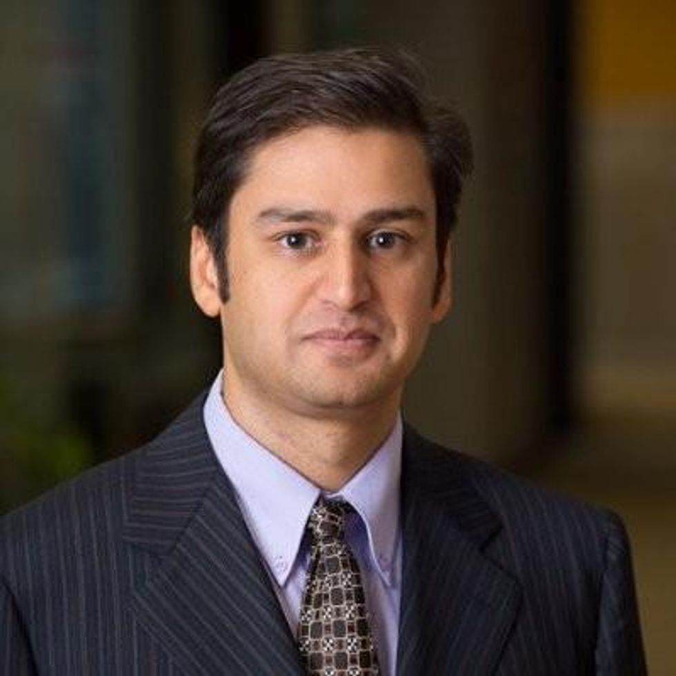
Dr. Amesh Adalja, Senior Scholar, Johns Hopkins Center for Health Security and an infectious disease physician
Listen to the Episode
Kira Peikoff was the editor-in-chief of Leaps.org from 2017 to 2021. As a journalist, her work has appeared in The New York Times, Newsweek, Nautilus, Popular Mechanics, The New York Academy of Sciences, and other outlets. She is also the author of four suspense novels that explore controversial issues arising from scientific innovation: Living Proof, No Time to Die, Die Again Tomorrow, and Mother Knows Best. Peikoff holds a B.A. in Journalism from New York University and an M.S. in Bioethics from Columbia University. She lives in New Jersey with her husband and two young sons. Follow her on Twitter @KiraPeikoff.

