Shoot for the Moon: Its Surface Contains a Pot of Gold

An astronaut standing on the Moon.
Here's a riddle: What do the Moon, nuclear weapons, clean energy of the future, terrorism, and lung disease all have in common?
One goal of India's upcoming space probe is to locate deposits of helium-3 that are worth trillions of dollars.
The answer is helium-3, a gas that's extremely rare on Earth but 100 million times more abundant on the Moon. This past October, the Lockheed Martin corporation announced a concept for a lunar landing craft that may return humans to the Moon in the coming decade, and yesterday China successfully landed the Change-4 probe on the far side of the Moon. Landing inside the Moon's deepest crater, the Chinese achieved a first in space exploration history.
Meanwhile, later this month, India's Chandrayaan-2 space probe will also land on the lunar surface. One of its goals is to locate deposits of helium-3 that are worth trillions of dollars, because it could be a fuel for nuclear fusion energy to generate electricity or propel a rocket.
The standard way that nuclear engineers are trying to achieve sustainable fusion uses fuels that are more plentiful on Earth: deuterium and tritium. But MIT researchers have found that adding small amounts of helium-3 to the mix could make it much more efficient, and thus a viable energy source much sooner that once thought.
Even if fusion is proven practical tomorrow, any kind of nuclear energy involves long waits for power plant construction measured in decades. However, mining helium-3 could be useful now, because of its non-energy applications. A major one is its ability to detect neutrons coming from plutonium that could be used in terrorist attacks. Here's how it works: a small amount of helium-3 is contained within a forensic instrument. When a neutron hits an atom of helium-3, the reaction produces tritium, a proton, and an electrical charge, alerting investigators to the possibility that plutonium is nearby.
Ironically, as global concern about a potential for hidden nuclear material increased in the early 2000s, so did the supply of helium-3 on Earth. That's because helium-3 comes from the decay of tritium, used in thermonuclear warheads (H-bombs). Thousands of such weapons have been dismantled from U.S. and Russian arsenals, making helium-3 available for plutonium detection, research, and other applications--including in the world of healthcare.
Helium-3 can help doctors diagnose lung diseases, since it enables imaging of the lungs in real time.
Helium-3 dramatically improves the ability of doctors to image the lungs in a range of diseases including asthma, chronic obstructive pulmonary disease and emphysema, cystic fibrosis, and bronchopulmonary dysplasia, which happens particularly in premature infants. Specifically, helium-3 is useful in magnetic resonance imaging (MRI), a procedure that creates images from within the body for diagnostic purposes.
But while a standard MRI allows doctors to visualize parts of the body like the heart or brain, it's useless for seeing the lungs. Because lungs are filled with air, which is much less dense than water or fat, effectively no signals are produced that would enable imaging.
To compensate for this problem, a patient can inhale gas that is hyperpolarized –meaning enhanced with special procedures so that the magnetic resonance signals from the lungs are finally readable. This gas is safe to breathe when mixed with enough oxygen to support life. Helium-3 is one such gas that can be hyperpolarized; since it produces such a strong signal, the MRI can literally see the air inside the lungs and in all of the airways, revealing intricate details of the bronchopulmonary tree. And it can do this in real time
The capability to show anatomic details of the lungs and airways, and the ability to display functional imaging as a patient breathes, makes helium-3 MRI far better than the standard method of testing lung function. Called spirometry, this method tells physicians how the lungs function overall, but does not home in on particular areas that may be causing a problem. Plus, spirometry requires patients to follow instructions and hold their breath, so it is not great for testing young children with pulmonary disease.
In recent years, the cost of helium-3 on Earth has skyrocketed.
Over the past several years, researchers have been developing MRI for lung testing using other hyperpolarized gases. The main alternative to helium-3 is xenon-129. Over the years, researchers have learned to overcome certain disadvantages of the latter, such as its potential to put patients to sleep. Since helium-3 provides the strongest signal, though, it is still the best gas for MRI studies in many lung conditions.
But the supply of helium-3 on Earth has been decreasing in recent years, due to the declining rate of dismantling of warheads, just as the Department of Homeland Security has required more and more of the gas for neutron detection. As a result, the cost of the gas has skyrocketed. Less is available now for medical uses – unless, of course, we begin mining it on the moon.
The question is: Are the benefits worth the 239,000-mile trip?
In this week's Friday Five, an old diabetes drug finds an exciting new purpose. Plus, how to make the cities of the future less toxic, making old mice younger with EVs, a new reason for mysterious stillbirths - and much more.
The Friday Five covers five stories in research that you may have missed this week. There are plenty of controversies and troubling ethical issues in science – and we get into many of them in our online magazine – but this news roundup focuses on scientific creativity and progress to give you a therapeutic dose of inspiration headed into the weekend.
Here is the promising research covered in this week's Friday Five:
Listen on Apple | Listen on Spotify | Listen on Stitcher | Listen on Amazon | Listen on Google
- How to make cities of the future less noisy
- An old diabetes drug could have a new purpose: treating an irregular heartbeat
- A new reason for mysterious stillbirths
- Making old mice younger with EVs
- No pain - or mucus - no gain
And an honorable mention this week: How treatments for depression can change the structure of the brain
Researchers at Stanford have found that the virus that causes Covid-19 can infect fat cells, which could help explain why obesity is linked to worse outcomes for those who catch Covid-19.
Obesity is a risk factor for worse outcomes for a variety of medical conditions ranging from cancer to Covid-19. Most experts attribute it simply to underlying low-grade inflammation and added weight that make breathing more difficult.
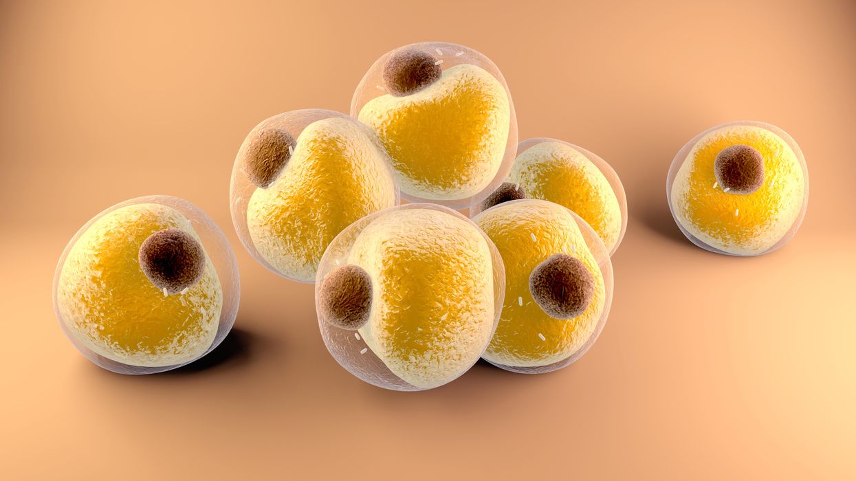
Now researchers have found a more direct reason: SARS-CoV-2, the virus that causes Covid-19, can infect adipocytes, more commonly known as fat cells, and macrophages, immune cells that are part of the broader matrix of cells that support fat tissue. Stanford University researchers Catherine Blish and Tracey McLaughlin are senior authors of the study.
Most of us think of fat as the spare tire that can accumulate around the middle as we age, but fat also is present closer to most internal organs. McLaughlin's research has focused on epicardial fat, “which sits right on top of the heart with no physical barrier at all,” she says. So if that fat got infected and inflamed, it might directly affect the heart.” That could help explain cardiovascular problems associated with Covid-19 infections.
Looking at tissue taken from autopsy, there was evidence of SARS-CoV-2 virus inside the fat cells as well as surrounding inflammation. In fat cells and immune cells harvested from health humans, infection in the laboratory drove "an inflammatory response, particularly in the macrophages…They secreted proteins that are typically seen in a cytokine storm” where the immune response runs amok with potential life-threatening consequences. This suggests to McLaughlin “that there could be a regional and even a systemic inflammatory response following infection in fat.”
It is easy to see how the airborne SARS-CoV-2 virus infects the nose and lungs, but how does it get into fat tissue? That is a mystery and the source of ample speculation.
The macrophages studied by McLaughlin and Blish were spewing out inflammatory proteins, While the the virus within them was replicating, the new viral particles were not able to replicate within those cells. It was a different story in the fat cells. “When [the virus] gets into the fat cells, it not only replicates, it's a productive infection, which means the resulting viral particles can infect another cell,” including microphages, McLaughlin explains. It seems to be a symbiotic tango of the virus between the two cell types that keeps the cycle going.
It is easy to see how the airborne SARS-CoV-2 virus infects the nose and lungs, but how does it get into fat tissue? That is a mystery and the source of ample speculation.
Macrophages are mobile; they engulf and carry invading pathogens to lymphoid tissue in the lymph nodes, tonsils and elsewhere in the body to alert T cells of the immune system to the pathogen. Perhaps some of them also carry the virus through the bloodstream to more distant tissue.
ACE2 receptors are the means by which SARS-CoV-2 latches on to and enters most cells. They are not thought to be common on fat cells, so initially most researchers thought it unlikely they would become infected.
However, while some cell receptors always sit on the surface of the cell, other receptors are expressed on the surface only under certain conditions. Philipp Scherer, a professor of internal medicine and director of the Touchstone Diabetes Center at the University of Texas Southwestern Medical Center, suggests that, in people who have obesity, “There might be higher levels of dysfunctional [fat cells] that facilitate entry of the virus,” either through transiently expressed ACE2 or other receptors. Inflammatory proteins generated by macrophages might contribute to this process.
Another hypothesis is that viral RNA might be smuggled into fat cells as cargo in small bits of material called extracellular vesicles, or EVs, that can travel between cells. Other researchers have shown that when EVs express ACE2 receptors, they can act as decoys for SARS-CoV-2, where the virus binds to them rather than a cell. These scientists are working to create drugs that mimic this decoy effect as an approach to therapy.
Do fat cells play a role in Long Covid? “Fat cells are a great place to hide. You have all the energy you need and fat cells turn over very slowly; they have a half-life of ten years,” says Scherer. Observational studies suggest that acute Covid-19 can trigger the onset of diabetes especially in people who are overweight, and that patients taking medicines to regulate their diabetes “were actually quite protective” against acute Covid-19. Scherer has funding to study the risks and benefits of those drugs in animal models of Long Covid.
McLaughlin says there are two areas of potential concern with fat tissue and Long Covid. One is that this tissue might serve as a “big reservoir where the virus continues to replicate and is sent out” to other parts of the body. The second is that inflammation due to infected fat cells and macrophages can result in fibrosis or scar tissue forming around organs, inhibiting their function. Once scar tissue forms, the tissue damage becomes more difficult to repair.
Current Covid-19 treatments work by stopping the virus from entering cells through the ACE2 receptor, so they likely would have no effect on virus that uses a different mechanism. That means another approach will have to be developed to complement the treatments we already have. So the best advice McLaughlin can offer today is to keep current on vaccinations and boosters and lose weight to reduce the risk associated with obesity.

