To Save Lives, This Scientist Is Trying to Grow Human Organs Inside of Sheep

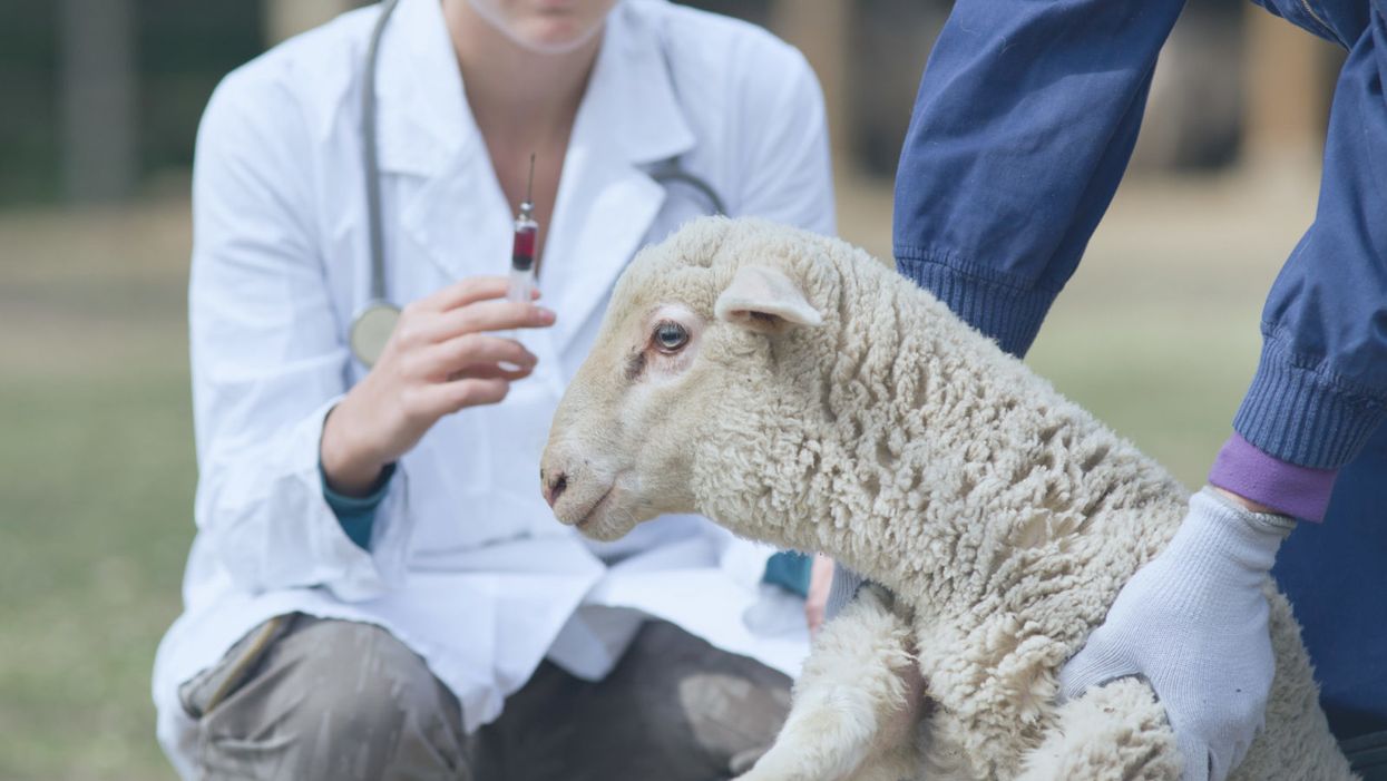
A lamb receiving a shot from medical personnel.
More than 114,000 men, women, and children are awaiting organ transplants in the United States. Each day, 22 of them die waiting. To address this shortage, researchers are working hard to grow organs on-demand, using the patient's own cells, to eliminate the need to find a perfectly matched donor.
"The next step is to transplant these cells into a larger animal that will produce an organ that is the right size for a human."
But creating full-size replacement organs in a lab is still decades away. So some scientists are experimenting with the boundaries of nature and life itself: using other mammals to grow human cells. Earlier this year, this line of investigation took a big step forward when scientists announced they had grown sheep embryos that contained human cells.
Dr. Pablo Ross, an associate professor at the University of California, Davis, along with a team of colleagues, introduced human stem cells into the sheep embryos at a very early stage of their development and found that one in every 10,000 cells in the embryo were human. It was an improvement over their prior experiment, using a pig embryo, when they found that one in every 100,000 cells in the pig were human. The resulting chimera, as the embryo is called, is only allowed to develop for 28 days. Leapsmag contributor Caren Chesler recently spoke with Ross about his research. Their interview has been edited and condensed for clarity.
Your goal is to one day grow human organs in animals, for organ transplantation. What does your research entail?
We're transplanting stem cells from a person into an animal embryo, at about day three to five of embryo development.
This concept has already been shown to work between mice and rats. You can grow a mouse pancreas inside a rat, or you can grow a rat pancreas inside a mouse.
For this approach to work for humans, the next step is to transplant these cells into a larger animal that will produce an organ that is the right size for a human. That's why we chose to start some of this preliminary work using pigs and sheep. Adult pigs and adult sheep have organs that are of similar size to an adult human. Pigs and sheep also grow really fast, so they can grow from a single cell at the time of fertilization to human adult size -- about 200 pounds -- in only nine to 10 months. That's better than the average waiting time for an organ transplant.
"You don't want the cells to confer any human characteristics in the animal....Too many cells, that may be a problem, because we do not know what that threshold is."
So how do you get the animal to grow the human organ you want?
First, we need to generate the animal without its own organ. We can generate sheep or pigs that will not grow their own pancreases. Those animals can then be used as hosts for human pancreas generation.
For the approach to work, we need the human stem cells to be able to integrate into the embryo and to contribute to its tissues. What we've been doing with pigs, and more recently, in sheep, is testing different types of stem cells, and introducing them into an early embryo between three to five days of development. We then transfer that embryo to a surrogate female and then harvest the embryos back at day 28 of development, at which point most of the organs are pre-formed.
The human cells will contribute to every organ. But in trying to do that, they will compete with the host organism. Since this is happening inside a pig embryo, which is inside a pig foster mother, the pig cells will win that competition for every organ.
Because you're not putting in enough human cells?
No, because it's a pig environment. Everything is pig. The host, basically, is in control. That's what we see when we do rat mice, or mouse rat: the host always wins the battle.
But we need human cells in the early development -- a few, but not too few -- so that when an organ needs to form, like a pancreas (which develops at around day 25), the pig cells will not respond to that, but if there are human cells in that location, [those human cells] can respond to pancreas formation.
From the work in mice and rats, we know we need some kind of global contribution across multiple tissues -- even a 1% contribution will be sufficient. But if the cells are not there, then they're not going to contribute to that organ. The way we target the specific organ is by removing the competition for that organ.
So if you want it to grow a pancreas, you use an embryo that is not going to grow a pancreas of its own. But you can't control where the other cells go. For instance, you don't want them going to the animal's brain – or its gonads –right?
You don't want the cells to confer any human characteristics in the animal. But even if cells go to the brain, it's not going to confer on the animal human characteristics. A few human cells, even if they're in the brain, won't make it a human brain. Too many cells, that may be a problem, because we do not know what that threshold is.
The objective of our research right now is to look at just 28 days of embryonic development and evaluate what's going on: Are the human cells there? How many? Do they go to the brain? If so, how many? Is this a problem, or is it not a problem? If we find that too many human cells go to the brain, that will probably mean that we wouldn't continue with this approach. At this point, we're not controlling it; we're analyzing it.
"By keeping our research in a very early stage of development, we're not creating a human or a humanoid or anything in between."
What other ethical concerns have arisen?
Conferring human properties to the organism, that is a major concern. I wouldn't like to be involved in that, and so that's what we're trying to assess. By keeping our research in a very early stage of development, we're not creating a human or a humanoid or anything in between.
What specifically sets off the ethical alarms? An animal developing human traits?
Animals developing human characteristics goes beyond what would be considered acceptable. I share that concern. But so far, what we have observed, primarily in rats and mice, is that the host animal dictates development. When you put mouse cells into a rat -- and they're so closely related, sometimes the mouse cells contribute to about 30 percent of the cells in the animal -- the outcome is still a rat. It's the size of a rat. It's the shape of the rat. It has the organ sizes of a rat. Even when the pancreas is fully made out of mouse cells, the pancreas is rat-sized because it grew inside the rat.
This happens even with an organ that is not shared, like a gallbladder, which mice have but rats do not. If you put cells from a mouse into a rat, it never grows a gallbladder. And if you put rat cells into the mouse, the rat cells can end up in the gallbladder even though those rat cells would never have made a gallbladder in a rat.
That means the cell structure is following the directions of the embryo, in terms of how they're going to form and what they're going to make. Based on those observations, if you put human cells into a sheep, we are going to get a sheep with human cells. The organs, the pancreas, in our case, will be the size and shape of the sheep pancreas, but it will be loaded with human cells identical to those of the patient that provided the cells used to generate the stem cells.
But, yeah, if by doing this, the animal acquires the functional or anatomical characteristics associated with a human, it would not be acceptable for me.
So you think these concerns are justified?
Absolutely. They need to be considered. But sometimes by raising these concerns, we prevent technologies from being developed. We need to consider the concerns, but we must evaluate them fully, to determine if they are scientifically justified. Because while we must consider the ethics of doing this, we also need to consider the ethics of not doing it. Every day, 22 people in the US die because they don't receive the organ they need to survive. This shortage is not going to be solved by donations, alone. That's clear. And when people die of old age, their organs are not good anymore.
Since organ transplantation has been so successful, the number of people needing organs has just been growing. The number of organs available has also grown but at a much slower pace. We need to find an alternative, and I think growing the organs in animals is one of those alternatives.
Right now, there's a moratorium on National Institutes of Health funding?
Yes. It's only one agency, but it happens to be the largest biomedical funding source. We have public funding for this work from the California Institute for Regenerative Medicine, and one of my colleagues has funding from the Department of Defense.
"I can say, without NIH funding, it's not going to happen here. It may happen in other places, like China."
Can we put the moratorium in context? How much research in the U.S. is funded by the NIH?
Probably more than 75 percent.
So what kind of impact would lifting that ban have on speeding up possible treatments for those who need a new organ?
Oh, I think it would have a huge impact. The moratorium not only prevents people from seeking funding to advance this area of research, it influences other sources of funding, who think, well, if the NIH isn't doing it, why are we going to do it? It hinders progress.
So with the ban, how long until we can really have organs growing in animals? I've heard five or 10 years.
With or without the ban, I don't think I can give you an accurate estimate.
What we know so far is that human cells don't contribute a lot to the animal embryo. We don't know exactly why. We have a lot of good ideas about things we can test, but we can't move forward right now because we don't have funding -- or we're moving forward but very slowly. We're really just scratching the surface in terms of developing these technologies.
We still need that one major leap in our understanding of how different species interact, and how human cells participate in the development of other species. I cannot predict when we're going to reach that point. I can say, without NIH funding, it's not going to happen here. It may happen in other places, like China, but without NIH funding, it's not going to happen in the U.S.
I think it's important to mention that this is in a very early stage of development and it should not be presented to people who need an organ as something that is possible right now. It's not fair to give false hope to people who are desperate.
So the five to 10 year figure is not realistic.
I think it will take longer than that. If we had a drug right now that we knew could stop heart attacks, it could take five to 10 years just to get it to market. With this, you're talking about a much more complex system. I would say 20 to 25 years. Maybe.
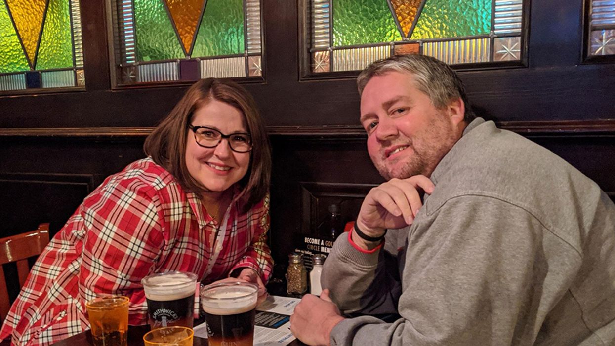
Jamie Rettinger with his now fiance Amie Purnel-Davis, who helped him through the clinical trial.
Jamie Rettinger was still in his thirties when he first noticed a tiny streak of brown running through the thumbnail of his right hand. It slowly grew wider and the skin underneath began to deteriorate before he went to a local dermatologist in 2013. The doctor thought it was a wart and tried scooping it out, treating the affected area for three years before finally removing the nail bed and sending it off to a pathology lab for analysis.
"I have some bad news for you; what we removed was a five-millimeter melanoma, a cancerous tumor that often spreads," Jamie recalls being told on his return visit. "I'd never heard of cancer coming through a thumbnail," he says. None of his doctors had ever mentioned it either. "I just thought I was being treated for a wart." But nothing was healing and it continued to bleed.
A few months later a surgeon amputated the top half of his thumb. Lymph node biopsy tested negative for spread of the cancer and when the bandages finally came off, Jamie thought his medical issues were resolved.
Melanoma is the deadliest form of skin cancer. About 85,000 people are diagnosed with it each year in the U.S. and more than 8,000 die of the cancer when it spreads to other parts of the body, according to the Centers for Disease Control and Prevention (CDC).
There are two peaks in diagnosis of melanoma; one is in younger women ages 30-40 and often is tied to past use of tanning beds; the second is older men 60+ and is related to outdoor activity from farming to sports. Light-skinned people have a twenty-times greater risk of melanoma than do people with dark skin.
"When I graduated from medical school, in 2005, melanoma was a death sentence" --Diwakar Davar.
Jamie had a follow up PET scan about six months after his surgery. A suspicious spot on his lung led to a biopsy that came back positive for melanoma. The cancer had spread. Treatment with a monoclonal antibody (nivolumab/Opdivo®) didn't prove effective and he was referred to the UPMC Hillman Cancer Center in Pittsburgh, a four-hour drive from his home in western Ohio.
An alternative monoclonal antibody treatment brought on such bad side effects, diarrhea as often as 15 times a day, that it took more than a week of hospitalization to stabilize his condition. The only options left were experimental approaches in clinical trials.
Early research
"When I graduated from medical school, in 2005, melanoma was a death sentence" with a cure rate in the single digits, says Diwakar Davar, 39, an oncologist at UPMC Hillman Cancer Center who specializes in skin cancer. That began to change in 2010 with introduction of the first immunotherapies, monoclonal antibodies, to treat cancer. The antibodies attach to PD-1, a receptor on the surface of T cells of the immune system and on cancer cells. Antibody treatment boosted the melanoma cure rate to about 30 percent. The search was on to understand why some people responded to these drugs and others did not.
At the same time, there was a growing understanding of the role that bacteria in the gut, the gut microbiome, plays in helping to train and maintain the function of the body's various immune cells. Perhaps the bacteria also plays a role in shaping the immune response to cancer therapy.
One clue came from genetically identical mice. Animals ordered from different suppliers sometimes responded differently to the experiments being performed. That difference was traced to different compositions of their gut microbiome; transferring the microbiome from one animal to another in a process known as fecal transplant (FMT) could change their responses to disease or treatment.
When researchers looked at humans, they found that the patients who responded well to immunotherapies had a gut microbiome that looked like healthy normal folks, but patients who didn't respond had missing or reduced strains of bacteria.
Davar and his team knew that FMT had a very successful cure rate in treating the gut dysbiosis of Clostridioides difficile, a persistant intestinal infection, and they wondered if a fecal transplant from a patient who had responded well to cancer immunotherapy treatment might improve the cure rate of patients who did not originally respond to immunotherapies for melanoma.

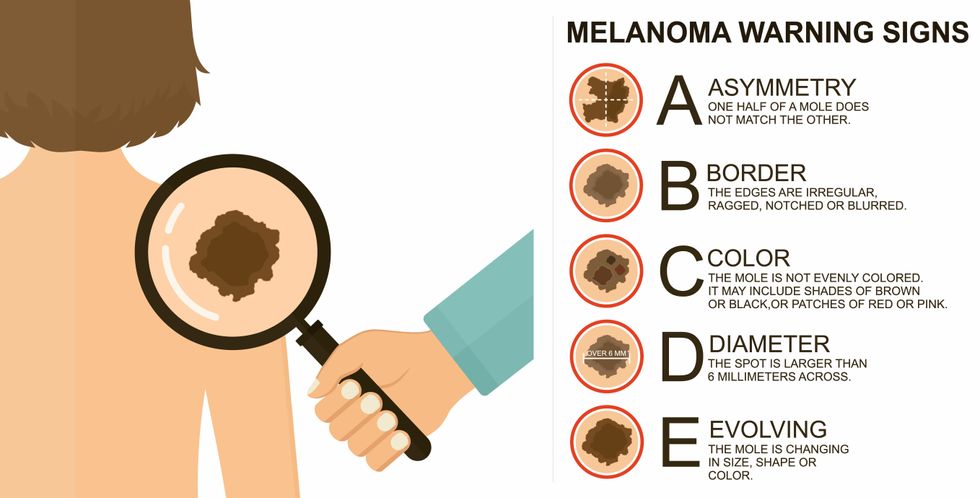
The ABCDE of melanoma detection
Adobe Stock
Clinical trial
"It was pretty weird, I was totally blasted away. Who had thought of this?" Jamie first thought when the hypothesis was explained to him. But Davar's explanation that the procedure might restore some of the beneficial bacterial his gut was lacking, convinced him to try. He quickly signed on in October 2018 to be the first person in the clinical trial.
Fecal donations go through the same safety procedures of screening for and inactivating diseases that are used in processing blood donations to make them safe for transfusion. The procedure itself uses a standard hollow colonoscope designed to screen for colon cancer and remove polyps. The transplant is inserted through the center of the flexible tube.
Most patients are sedated for procedures that use a colonoscope but Jamie doesn't respond to those drugs: "You can't knock me out. I was watching them on the TV going up my own butt. It was kind of unreal at that point," he says. "There were about twelve people in there watching because no one had seen this done before."
A test two weeks after the procedure showed that the FMT had engrafted and the once-missing bacteria were thriving in his gut. More importantly, his body was responding to another monoclonal antibody (pembrolizumab/Keytruda®) and signs of melanoma began to shrink. Every three months he made the four-hour drive from home to Pittsburgh for six rounds of treatment with the antibody drug.
"We were very, very lucky that the first patient had a great response," says Davar. "It allowed us to believe that even though we failed with the next six, we were on the right track. We just needed to tweak the [fecal] cocktail a little better" and enroll patients in the study who had less aggressive tumor growth and were likely to live long enough to complete the extensive rounds of therapy. Six of 15 patients responded positively in the pilot clinical trial that was published in the journal Science.
Davar believes they are beginning to understand the biological mechanisms of why some patients initially do not respond to immunotherapy but later can with a FMT. It is tied to the background level of inflammation produced by the interaction between the microbiome and the immune system. That paper is not yet published.
Surviving cancer
It has been almost a year since the last in his series of cancer treatments and Jamie has no measurable disease. He is cautiously optimistic that his cancer is not simply in remission but is gone for good. "I'm still scared every time I get my scans, because you don't know whether it is going to come back or not. And to realize that it is something that is totally out of my control."
"It was hard for me to regain trust" after being misdiagnosed and mistreated by several doctors he says. But his experience at Hillman helped to restore that trust "because they were interested in me, not just fixing the problem."
He is grateful for the support provided by family and friends over the last eight years. After a pause and a sigh, the ruggedly built 47-year-old says, "If everyone else was dead in my family, I probably wouldn't have been able to do it."
"I never hesitated to ask a question and I never hesitated to get a second opinion." But Jamie acknowledges the experience has made him more aware of the need for regular preventive medical care and a primary care physician. That person might have caught his melanoma at an earlier stage when it was easier to treat.
Davar continues to work on clinical studies to optimize this treatment approach. Perhaps down the road, screening the microbiome will be standard for melanoma and other cancers prior to using immunotherapies, and the FMT will be as simple as swallowing a handful of freeze-dried capsules off the shelf rather than through a colonoscopy. Earlier this year, the Food and Drug Administration approved the first oral fecal microbiota product for C. difficile, hopefully paving the way for more.
An older version of this hit article was first published on May 18, 2021
All organisms can repair damaged tissue, but none do it better than salamanders and newts. A surprising area of science could tell us how they manage this feat - and perhaps even help us develop a similar ability.
All organisms have the capacity to repair or regenerate tissue damage. None can do it better than salamanders or newts, which can regenerate an entire severed limb.
That feat has amazed and delighted man from the dawn of time and led to endless attempts to understand how it happens – and whether we can control it for our own purposes. An exciting new clue toward that understanding has come from a surprising source: research on the decline of cells, called cellular senescence.
Senescence is the last stage in the life of a cell. Whereas some cells simply break up or wither and die off, others transition into a zombie-like state where they can no longer divide. In this liminal phase, the cell still pumps out many different molecules that can affect its neighbors and cause low grade inflammation. Senescence is associated with many of the declining biological functions that characterize aging, such as inflammation and genomic instability.
Oddly enough, newts are one of the few species that do not accumulate senescent cells as they age, according to research over several years by Maximina Yun. A research group leader at the Center for Regenerative Therapies Dresden and the Max Planck Institute of Molecular and Cell Biology and Genetics, in Dresden, Germany, Yun discovered that senescent cells were induced at some stages of regeneration of the salamander limb, “and then, as the regeneration progresses, they disappeared, they were eliminated by the immune system,” she says. “They were present at particular times and then they disappeared.”
Senescent cells added to the edges of the wound helped the healthy muscle cells to “dedifferentiate,” essentially turning back the developmental clock of those cells into more primitive states.
Previous research on senescence in aging had suggested, logically enough, that applying those cells to the stump of a newly severed salamander limb would slow or even stop its regeneration. But Yun stood that idea on its head. She theorized that senescent cells might also play a role in newt limb regeneration, and she tested it by both adding and removing senescent cells from her animals. It turned out she was right, as the newt limbs grew back faster than normal when more senescent cells were included.
Senescent cells added to the edges of the wound helped the healthy muscle cells to “dedifferentiate,” essentially turning back the developmental clock of those cells into more primitive states, which could then be turned into progenitors, a cell type in between stem cells and specialized cells, needed to regrow the muscle tissue of the missing limb. “We think that this ability to dedifferentiate is intrinsically a big part of why salamanders can regenerate all these very complex structures, which other organisms cannot,” she explains.
Yun sees regeneration as a two part problem. First, the cells must be able to sense that their neighbors from the lost limb are not there anymore. Second, they need to be able to produce the intermediary progenitors for regeneration, , to form what is missing. “Molecularly, that must be encoded like a 3D map,” she says, otherwise the new tissue might grow back as a blob, or liver, or fin instead of a limb.
Wound healing
Another recent study, this time at the Mayo Clinic, provides evidence supporting the role of senescent cells in regeneration. Looking closely at molecules that send information between cells in the wound of a mouse, the researchers found that senescent cells appeared near the start of the healing process and then disappeared as healing progressed. In contrast, persistent senescent cells were the hallmark of a chronic wound that did not heal properly. The function and significance of senescence cells depended on both the timing and the context of their environment.
The paper suggests that senescent cells are not all the same. That has become clearer as researchers have been able to identify protein markers on the surface of some senescent cells. The patterns of these proteins differ for some senescent cells compared to others. In biology, such physical differences suggest functional differences, so it is becoming increasingly likely there are subsets of senescent cells with differing functions that have not yet been identified.
There are disagreements within the research community as to whether newts have acquired their regenerative capacity through a unique evolutionary change, or if other animals, including humans, retain this capacity buried somewhere in their genes.
Scientists initially thought that senescent cells couldn’t play a role in regeneration because they could no longer reproduce, says Anthony Atala, a practicing surgeon and bioengineer who leads the Wake Forest Institute for Regenerative Medicine in North Carolina. But Yun’s study points in the other direction. “What this paper shows clearly is that these cells have the potential to be involved in tissue regeneration [in newts]. The question becomes, will these cells be able to do the same in humans.”
As our knowledge of senescent cells increases, Atala thinks we need to embrace a new analogy to help understand them: humans in retirement. They “have acquired a lot of wisdom throughout their whole life and they can help younger people and mentor them to grow to their full potential. We're seeing the same thing with these cells,” he says. They are no longer putting energy into their own reproduction, but the signaling molecules they secrete “can help other cells around them to regenerate.”
There are disagreements within the research community as to whether newts have acquired their regenerative capacity through a unique evolutionary change, or if other animals, including humans, retain this capacity buried somewhere in their genes. If so, it seems that our genes are unable to express this ability, perhaps as part of a tradeoff in acquiring other traits. It is a fertile area of research.
Dedifferentiation is likely to become an important process in the field of regenerative medicine. One extreme example: a lab has been able to turn back the clock and reprogram adult male skin cells into female eggs, a potential milestone in reproductive health. It will be more difficult to control just how far back one wishes to go in the cell's dedifferentiation – part way or all the way back into a stem cell – and then direct it down a different developmental pathway. Yun is optimistic we can learn these tricks from newts.
Senolytics
A growing field of research is using drugs called senolytics to remove senescent cells and slow or even reverse disease of aging.
“Senolytics are great, but senolytics target different types of senescence,” Yun says. “If senescent cells have positive effects in the context of regeneration, of wound healing, then maybe at the beginning of the regeneration process, you may not want to take them out for a little while.”
“If you look at pretty much all biological systems, too little or too much of something can be bad, you have to be in that central zone” and at the proper time, says Atala. “That's true for proteins, sugars, and the drugs that you take. I think the same thing is true for these cells. Why would they be different?”
Our growing understanding that senescence is not a single thing but a variety of things likely means that effective senolytic drugs will not resemble a single sledge hammer but more a carefully manipulated scalpel where some types of senescent cells are removed while others are added. Combinations and timing could be crucial, meaning the difference between regenerating healthy tissue, a scar, or worse.

