

Often called the window to the soul, the eyes are more sacred than other body parts, at least for some.
Eye transplants are desperately needed, but they're nowhere in sight. About 12.7 million people worldwide need a corneal transplant, which means that only one in 70 people who require them, get them. The gaps are international. Eye banks in the United Kingdom are around 20 percent below the level needed to supply hospitals, while Indian eye banks, which need at least 250,000 corneas per year, collect only around 45 to 50 thousand donor corneas (and of those 60 to 70 percent are successfully transplanted).
As for retinas, it's impossible currently to put one into the eye of another person. Artificial devices can be implanted to restore the sight of patients suffering from severe retinal diseases, but the number of people around the world with such “bionic eyes” is less than 600, while in America alone 11 million people have some type of retinal disease leading to severe vision loss. Add to this an increasingly aging population, commonly facing various vision impairments, and you have a recipe for heavy burdens on individuals, the economy and society. In the U.S. alone, the total annual economic impact of vision problems was $51.4 billion in 2017.
Even if you try growing tissues in the petri dish route into organoids mimicking the function of the human eye, you will not get the physiological complexity of the structure and metabolism of the real thing, according to Cosma. She is a member of a scientific consortium that includes researchers from major institutions from Spain, the U.K., Portugal, Italy and Israel. The consortium has received about $3.8 million from the European Union to pursue innovative eye research. Her team’s goal is to give hope to at least 2.2 billion people across the world afflicted with a vision impairment and 33 million who go through life with avoidable blindness.
Their method? Resuscitating cadaveric eyes for at least a month.
If we succeed, it will be the first intact human model of the eye capable of exploring and analyzing regenerative processes ex vivo. -- Maria Pia Cosma.
“We proposed to resuscitate eyes, that is to restore the global physiology and function of human explanted tissues,” Cosma said, referring to living tissues extracted from the eye and placed in a medium for culture. Their ECaBox is an ex vivo biological system, in which eyes taken from dead donors are placed in an artificial environment, designed to preserve the eye’s temperature and pH levels, deter blood clots, and remove the metabolic waste and toxins that would otherwise spell their demise.
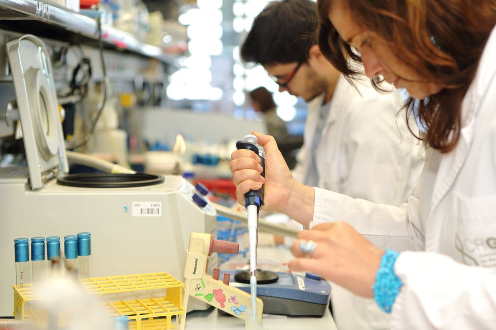
Scientists work on resuscitating eyes in the lab of Maria Pia Cosma.
Courtesy of Maria Pia Cosma.
“One of the great challenges is the passage of the blood in the capillary branches of the eye, what we call long-term perfusion,” Cosma said. Capillaries are an intricate network of very thin blood vessels that transport blood, nutrients and oxygen to cells in the body’s organs and systems. To maintain the garland-shaped structure of this network, sufficient amounts of oxygen and nutrients must be provided through the eye circulation and microcirculation. “Our ambition is to combine perfusion of the vessels with artificial blood," along with using a synthetic form of vitreous, or the gel-like fluid that lets in light and supports the the eye's round shape, Cosma said.
The scientists use this novel setup with the eye submersed in its medium to keep the organ viable, so they can test retinal function. “If we succeed, we will ensure full functionality of a human organ ex vivo. It will be the first intact human model of the eye capable of exploring and analyzing regenerative processes ex vivo,” Cosma added.
A rapidly developing field of regenerative medicine aims to stimulate the body's natural healing processes and restore or replace damaged tissues and organs. But for people with retinal diseases, regenerative medicine progress has been painfully slow. “Experiments on rodents show progress, but the risks for humans are unacceptable,” Cosma said.
The ECaBox could boost progress with regenerative medicine for people with retinal diseases, which has been painfully slow because human experiments involving their eyes are too risky. “We will test emerging treatments while reducing animal research, and greatly accelerate the discovery and preclinical research phase of new possible treatments for vision loss at significantly reduced costs,” Cosma explained. Much less time and money would be wasted during the drug discovery process. Their work may even make it possible to transplant the entire eyeball for those who need it.
“It is a very exciting project,” said Sanjay Sharma, a professor of ophthalmology and epidemiology at Queen's University, in Kingston, Canada. “The ability to explore and monitor regenerative interventions will increasingly be of importance as we develop therapies that can regenerate ocular tissues, including the retina.”
Seemingly, there's no sacred religious text or a holy book prohibiting the practice of eye donation.
But is the world ready for eye transplants? “People are a bit weird or very emotional about donating their eyes as compared to other organs,” Cosma said. And much can be said about the problem of eye donor shortage. Concerns include disfigurement and healthcare professionals’ fear that the conversation about eye donation will upset the departed person’s relatives because of cultural or religious considerations. As just one example, Sharma noted the paucity of eye donations in his home country, Canada.
Yet, experts like Sharma stress the importance of these donations for both the recipients and their family members. “It allows them some psychological benefit in a very difficult time,” he said. So why are global eye banks suffering? Is it because the eyes are the windows to the soul?
Seemingly, there's no sacred religious text or a holy book prohibiting the practice of eye donation. In fact, most major religions of the world permit and support organ transplantation and donation, and by extension eye donation, because they unequivocally see it as an “act of neighborly love and charity.” In Hinduism, the concept of eye donation aligns with the Hindu principle of daan or selfless giving, where individuals donate their organs or body after death to benefit others and contribute to society. In Islam, eye donation is a form of sadaqah jariyah, a perpetual charity, as it can continue to benefit others even after the donor's death.
Meanwhile, Buddhist masters teach that donating an organ gives another person the chance to live longer and practice dharma, the universal law and order, more meaningfully; they also dismiss misunderstandings of the type “if you donate an eye, you’ll be born without an eye in the next birth.” And Christian teachings emphasize the values of love, compassion, and selflessness, all compatible with organ donation, eye donation notwithstanding; besides, those that will have a house in heaven, will get a whole new body without imperfections and limitations.
The explanation for people’s resistance may lie in what Deepak Sarma, a professor of Indian religions and philosophy at Case Western Reserve University in Cleveland, calls “street interpretation” of religious or spiritual dogmas. Consider the mechanism of karma, which is about the causal relation between previous and current actions. “Maybe some Hindus believe there is karma in the eyes and, if the eye gets transplanted into another person, they will have to have that karmic card from now on,” Sarma said. “Even if there is peculiar karma due to an untimely death–which might be interpreted by some as bad karma–then you have the karma of the recipient, which is tremendously good karma, because they have access to these body parts, a tremendous gift,” Sarma said. The overall accumulation is that of good karma: “It’s a beautiful kind of balance,” Sarma said.
For the Jews, Christians, and Muslims who believe in the physical resurrection of the body that will be made new in an afterlife, the already existing body is sacred since it will be the basis of a new refashioned body in an afterlife.---Omar Sultan Haque.
With that said, Sarma believes it is a fallacy to personify or anthropomorphize the eye, which doesn’t have a soul, and stresses that the karma attaches itself to the soul and not the body parts. But for scholars like Omar Sultan Haque—a psychiatrist and social scientist at Harvard Medical School, investigating questions across global health, anthropology, social psychology, and bioethics—the hierarchy of sacredness of body parts is entrenched in human psychology. You cannot equate the pinky toe with the face, he explained.
“The eyes are the window to the soul,” Haque said. “People have a hierarchy of body parts that are considered more sacred or essential to the self or soul, such as the eyes, face, and brain.” In his view, the techno-utopian transhumanist communities (especially those in Silicon Valley) have reduced the totality of a person to a mere material object, a “wet robot” that knows no sacredness or hierarchy of human body parts. “But for the Jews, Christians, and Muslims who believe in the physical resurrection of the body that will be made new in an afterlife, the [already existing] body is sacred since it will be the basis of a new refashioned body in an afterlife,” Haque said. “You cannot treat the body like any old material artifact, or old chair or ragged cloth, just because materialistic, secular ideologies want so,” he continued.
For Cosma and her peers, however, the very definition of what is alive or not is a bit semantic. “As soon as we die, the electrophysiological activity in the eye stops,” she said. “The goal of the project is to restore this activity as soon as possible before the highly complex tissue of the eye starts degrading.” Cosma’s group doesn’t yet know when they will be able to keep the eyes alive and well in the ECaBox, but the consensus is that the sooner the better. Hopefully, the taboos and fears around the eye donations will dissipate around the same time.

When graduating college this month, many North American engineering students will take a special pledge, with a history dating back to 1925.
The tradition of taking an engineering oath began over a century ago in Canada. In 1922, Herbert E.T. Haultain, professor of mining engineering at the University of Toronto, presented the idea at the annual meeting of the Engineering Institute of Canada. The seven past presidents of that body were in attendance, heard Haultain’s speech and accepted his suggestion to form a committee to create an honor oath. Later, they formed the nonprofit Corporation of the Seven Wardens, which would oversee the ritual. Next year, in 1923, with the encouragement of the Seven Wardens, Haultain wrote to poet and writer Rudyard Kipling, asking him to develop a professional oath for engineers. “We are a tribe—a very important tribe within the community,” Haultain said in the letter, “but we are lacking in tribal spirit, or perhaps I should say, in manifestation of tribal spirit. Also, we are inarticulate. Can you help us?”
While Kipling is most famous now for “The Jungle Book” and perhaps his poem “Gunga Din,” he had also written a short story about engineers, “The Bridge Builders.” His poem “The Sons of Martha” can be read as a celebration of engineers:
It is their care in all the ages to take the buffet and cushion the shock.
It is their care that the gear engages; it is their care that the switches lock.
It is their care that the wheels run truly; it is their care to embark and entrain,
Tally, transport, and deliver duly the Sons of Mary by land and main.
Kipling accepted the ask and wrote the Ritual of the Calling of an Engineer, which he sent to Haultain a month later. In his response to Haultain, he stated that he preferred the word “Obligation” to “Oath.” He wrote the Obligation using Old English lettering and the old-fashioned capitalization. Kipling’s Obligation binds engineers upon their “Honor and Cold Iron” to not “suffer or pass, or be privy to the passing of, Bad Workmanship or Faulty Material,” and pardon is asked “in the presence of my betters and my equals in my Calling” for the engineer’s “assured failures and derelictions.” The hope is that when one is tempted to shoddy work by weakness or weariness, the memory of the Obligation “and the company before whom it was entered into, may return to me to aid, comfort, and restrain.”
Using the Obligation, The Seven Wardens created an induction ceremony, which seeks to unify the profession and recognize engineering’s ethics, including responsibility to the public and the need to make the best decisions possible. The induction ceremony included recitation of Kipling’s “Obligation” and incorporated an anvil, a hammer, an iron chain, and an iron ring. The inductee engineers sat inside an area marked off by the iron chain, with their more senior colleagues outside that area. At the start of the ritual, the leader beat out S-S-T in Morse code with the hammer and anvil—the letters standing for Steel, Stone, and Time. A more experienced and previously obligated engineer placed the ring on the small finger of the inductee engineer’s working hand. As per Kipling, the ring’s rough, faceted texture symbolized “the young engineer’s mind” and the difficulties engineers face in mastering their discipline.
A persistent myth purports that the original iron rings were made from the beams or bolts of the Quebec Bridge that failed twice during construction.
The first induction ceremony took place on April 25, 1925, in Montreal to obligate two of the Seven Wardens, along with four graduates from the University of Toronto class of 1893. On May 1 of that year, 14 more engineers were obligated at the University of Toronto. From that time to today most Canadian professional engineers have gone through that same ritual in their various camps, called Kipling camps—local chapters associated with various Canadian universities.
Henry Petroski, Duke University’s professor of civil engineering and history, notes in his book, “Forgive Design: Understanding Failure,” that Kipling’s poem “Sons of Martha” is often read as part of the ritual. However, sometimes inductees read Kipling’s “Hymn of Breaking Strain,” instead, which graphically depicts disastrous outcomes of engineering mistakes. The first stanza of that poem says:
The careful text-books measure
(Let all who build beware!)
The load, the shock, the pressure
Material can bear.
So, when the buckled girder
Lets down the grinding span,
'The blame of loss, or murder,
Is laid upon the man.
Not on the Stuff—the Man!
As if to strengthen the importance of these concepts, a persistent myth purports that the original iron rings were made from the beams or bolts of the Quebec Bridge that failed twice during construction. The bridge spans the St. Lawrence River upriver from Quebec City, and at the time of its construction was the world’s longest at 1,800 feet. Due to engineering errors and poor oversight, the bridge’s own weight exceeded its carrying capacity. Moreover, engineers downplayed danger when bridge beams began to warp under stress, saying that they were probably warped before they were installed. On August 29, 1907, the bridge collapsed, killing 75 of 86 workers. A second collapse occurred in 1916 when lifting equipment failed, and thirteen more workers died.
The ring myth, however, couldn’t be true. The original iron rings couldn’t have come from the failed bridge since it was made of steel, not wrought iron. Today the rings are made from stainless steel because iron deteriorates and stains engineers’ finger black.

On August 14, 2018, Morandi Bridge over Polcevera River in Genoa, Italy, collapsed from structural failure, killing 43 people.
Adobe Stock
The Seven Wardens decided to restrict the ritual to engineers trained in Canada. They copyrighted the obligation oath in Canada and the United States in 1935. Although the ritual is not a requirement for professional licensing, just like the Hippocratic Oath is not part of medical licensing, it remains a long-standing tradition.
The American Obligation of the Engineer has its own creation story, albeit a very different one. The American Order of the Engineer (OOE) was initiated in 1970, during the era of the anti-war protests, Apollo missions and the first Earth Day. On May 4, 1970, the National Guard shot into a crowd of protesters at Kent State University, killing four people. The two authors of the American obligation—Cleveland State University’s (CSU) engineering professor John Janssen and his wife Susan—reflected these historical events in the oath they wrote. Their version of the oath binds engineers to “practice integrity and fair dealing.” It also notes that their “skill carries with it the obligation to serve humanity by making the best use of the Earth’s precious wealth.” As Petroski explains in his book, “campus antiwar protestors around the country tended to view engineers as complicit in weapons proliferation [which] prompted some [CSU] engineering student leaders to look for a means of asserting some more positive values.”
Kip A. Wedel, associate professor of history and politics at Bethel College, wrote in his book, “The Obligation: A History of the Order of the Engineer,” that the ceremony was not a direct response to the Kent State shootings—it was already scheduled when the shootings happened. Yet, engineering students found the ceremony a positive action they could take in contrast to the overall turmoil. The first American ritual took place on June 4, 1970, at CSU. In total, 170 students, faculty members, and practicing engineers took the obligation. This established CSU as the first Link of the Order, as the OOE designates its local chapters. For their first ceremony, the CSU students fabricated smooth, unfaceted rings from stainless steel pipe. Later they were replaced by factory-made rings. According to Paula Ostaff, OOE’s Executive Director, about 20,000 eligible students and alumni obligate themselves yearly.
Societies hope that every engineer is imbued with a strong ethical sense and that their pledges are never far from mind. For some, the rings they wear serve a daily reminder that every paper they sign off on is touched by a physical reminder of their commitment.
These ethical and responsible engineering practices are especially salient today, when one in three American bridges needs repair or replacement, some have already collapsed, and engineers are working on projects related to the bipartisan infrastructure bill President Biden signed into law in 2021. Canada has committed $33 billion to its Investing in Canada Infrastructure Program. At the heart of these grand projects are many thousands of professional engineers, collectively working millions of hours. The professional vows they took aim to assure that the homes, bridges and airplanes they build will work as expected.
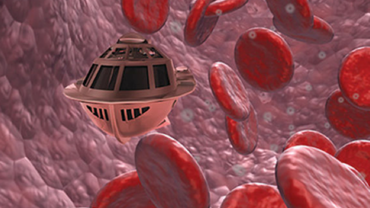
A movie still from the 1966 film "Fantastic Voyage"
"Chemotherapy is delivered systemically," Bionaut-founder and CEO Michael Shpigelmacher says. "Often only a small percentage arrives at the location where it is actually needed."
But what if it was possible to send a tiny robot through the body to attack a tumor or deliver a drug at exactly the right location?
Several startups and academic institutes worldwide are working to develop such a solution but Bionaut Labs seems the furthest along in advancing its invention. "You can think of the Bionaut as a tiny screw that moves through the veins as if steered by an invisible screwdriver until it arrives at the tumor," Shpigelmacher explains. Via Zoom, he shares the screen of an X-ray machine in his Culver City lab to demonstrate how the half-transparent, yellowish device winds its way along the spine in the body. The nanobot contains a tiny but powerful magnet. The "invisible screwdriver" is an external magnetic field that rotates that magnet inside the device and gets it to move and change directions.
The current model has a diameter of less than a millimeter. Shpigelmacher's engineers could build the miniature vehicle even smaller but the current size has the advantage of being big enough to see with bare eyes. It can also deliver more medicine than a tinier version. In the Zoom demonstration, the micorobot is injected into the spine, not unlike an epidural, and pulled along the spine through an outside magnet until the Bionaut reaches the brainstem. Depending which organ it needs to reach, it could be inserted elsewhere, for instance through a catheter.
"The hope is that we can develop a vehicle to transport medication deep into the body," says Max Planck scientist Tian Qiu.
Imagine moving a screw through a steak with a magnet — that's essentially how the device works. But of course, the Bionaut is considerably different from an ordinary screw: "At the right location, we give a magnetic signal, and it unloads its medicine package," Shpigelmacher says.
To start, Bionaut Labs wants to use its device to treat Parkinson's disease and brain stem gliomas, a type of cancer that largely affects children and teenagers. About 300 to 400 young people a year are diagnosed with this type of tumor. Radiation and brain surgery risk damaging sensitive brain tissue, and chemotherapy often doesn't work. Most children with these tumors live less than 18 months. A nanobot delivering targeted chemotherapy could be a gamechanger. "These patients really don't have any other hope," Shpigelmacher says.
Of course, the main challenge of the developing such a device is guaranteeing that it's safe. Because tissue is so sensitive, any mistake could risk disastrous results. In recent years, Bionaut has tested its technology in dozens of healthy sheep and pigs with no major adverse effects. Sheep make a good stand-in for humans because their brains and spines are similar to ours.
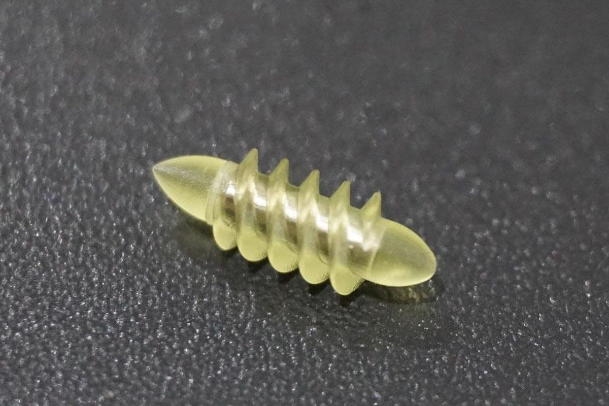
The Bionaut device is about the size of a grain of rice.
Bionaut Labs
"As the Bionaut moves through brain tissue, it creates a transient track that heals within a few weeks," Shpigelmacher says. The company is hoping to be the first to test a nanobot in humans. In December 2022, it announced that a recent round of funding drew $43.2 million, for a total of 63.2 million, enabling more research and, if all goes smoothly, human clinical trials by early next year.
Once the technique has been perfected, further applications could include addressing other kinds of brain disorders that are considered incurable now, such as Alzheimer's or Huntington's disease. "Microrobots could serve as a bridgehead, opening the gateway to the brain and facilitating precise access of deep brain structure – either to deliver medication, take cell samples or stimulate specific brain regions," Shpigelmacher says.
Robot-assisted hybrid surgery with artificial intelligence is already used in state-of-the-art surgery centers, and many medical experts believe that nanorobotics will be the instrument of the future. In 2016, three scientists were awarded the Nobel Prize in Chemistry for their development of "the world's smallest machines," nano "elevators" and minuscule motors. Since then, the scientific experiments have progressed to the point where applicable devices are moving closer to actually being implemented.
Bionaut's technology was initially developed by a research team lead by Peer Fischer, head of the independent Micro Nano and Molecular Systems Lab at the Max Planck Institute for Intelligent Systems in Stuttgart, Germany. Fischer is considered a pioneer in the research of nano systems, which he began at Harvard University more than a decade ago. He and his team are advising Bionaut Labs and have licensed their technology to the company.
"The hope is that we can develop a vehicle to transport medication deep into the body," says Max Planck scientist Tian Qiu, who leads the cooperation with Bionaut Labs. He agrees with Shpigelmacher that the Bionaut's size is perfect for transporting medication loads and is researching potential applications for even smaller nanorobots, especially in the eye, where the tissue is extremely sensitive. "Nanorobots can sneak through very fine tissue without causing damage."
In "Fantastic Voyage," Raquel Welch's adventures inside the body of a dissident scientist let her swim through his veins into his brain, but her shrunken miniature submarine is attacked by antibodies; she has to flee through the nerves into the scientist's eye where she escapes into freedom on a tear drop. In reality, the exit in the lab is much more mundane. The Bionaut simply leaves the body through the same port where it entered. But apart from the dramatization, the "Fantastic Voyage" was almost prophetic, or, as Shpigelmacher says, "Science fiction becomes science reality."
This article was first published by Leaps.org on April 12, 2021.