CRISPR base editing gives measure of hope to people with muscular dystrophy

Next year, a neurologist will test CRISPR base editing in a trial of five people with muscular dystrophy to see if their muscles accept corrected cells and whether they multiply and take over the function of damaged cells.
When Martin Weber climbs the steps to his apartment on the fifth floor in Munich, an attentive observer might notice that he walks a little unevenly. “That’s because my calf muscles were the first to lose strength,” Weber explains.
About three years ago, the now 19-year-old university student realized that he suddenly had trouble keeping up with his track team at school. At tennis tournaments, he seemed to lose stamina after the first hour. “But it was still within the norm,” he says. “So it took a while before I noticed something was seriously wrong.” A blood test showed highly elevated liver markers. His parents feared he had liver cancer until a week-long hospital visit and scores of tests led to a diagnosis: hereditary limb-girdle muscular dystrophy, an incurable genetic illness that causes muscles to deteriorate.
As you read this text, you will surely use several muscles without being aware of them: Your heart muscle pumps blood through your arteries, your eye muscles let you follow the words in this sentence, and your hand muscles hold the tablet or cell phone. Muscles make up 40 percent of your body weight; we usually have 656 of them. Now imagine they are slowly losing their strength. No training, no protein shake can rebuild their function.
This is the reality for most people in Simone Spuler’s outpatient clinic at the Charité Hospital in Berlin, Germany: Almost all of her 2,500 patients have muscular dystrophy, a progressive illness striking mostly young people. Muscle decline leads to a wheelchair and, eventually, an early death due to a heart attack or the inability to breathe. In Germany alone, 300,000 people live with this illness, the youngest barely a year old. The CDC estimates that its most common form, Duchenne, affects 1 in every 3,500 to 6,000 male births each year in the United States.
The devastating progression of the disease is what motivates Spuler and her team of 25 scientists to find a cure. In 2019, they made a spectacular breakthrough: For the first time, they successfully used mRNA to introduce the CRISPR-Cas9 tool into human muscle stem cells to repair the dystrophy. “It’s really just one tiny molecule that doesn’t work properly,” Spuler explains.
CRISPR-Cas9 is a technology that lets scientists select and alter parts of the genome. It’s still comparatively new but has advanced quickly since its discovery in the early 2010s. “We now have the possibility to repair certain mutations with genetic editing,” Spuler says. “It’s pure magic.”
She projects a warm, motherly air and a professional calm that inspires trust from her patients. She needs these qualities because the 60-year-old neurologist has one of the toughest jobs in the world: All day long, patients with the incurable diagnosis of muscular dystrophy come to her clinic, and she watches them decline over the years. “Apart from physiotherapy, there is nothing we can recommend right now,” she says. That motivated her early in her career, when she met her first patients at the Max Planck Institute for Neurobiology near Munich in the 1990s. “I knew I had 30, 40 years to find something.”
She learned from the luminaries of her profession with postdocs at the University of California San Diego, Harvard and Johns Hopkins, before serving as a clinical fellow at the Mayo Clinic. In 2005, the Charité offered her the opportunity to establish a specialized clinic for myasthenia, or muscular weakness. An important influence on Spuler, she says, has been the French microbiologist Emmanuelle Charpentier, who received the Nobel Prize in 2020 along with Jennifer Doudna for their CRISPR research, and has worked in Berlin since 2015.
When CRISPR was first introduced, it was mainly used to cut through DNA. However, the cut can lead to undesired side effects. For the muscle stem cells, Spuler now uses a base editor to repair the damaged molecule with super fine scissors or tweezers.

“Apart from physiotherapy, there is nothing we can recommend right now,” Spuler says about her patients with limb-girdle muscular dystrophy.
Pablo Castagnola
Last year, she proved that the method works in mice. Injecting repaired cells into the rodents led to new muscle fibers and, in 2021 and 2022, she passed the first safety meetings with the Paul-Ehrlich Institute, which is responsible for approving human gene editing trials in Germany. She raised the nearly four million Euros needed to test the new method in the first clinical trial in humans with limb-girdle muscular dystrophy, beginning with one muscle that can easily be measured, such as the biceps.
This spring, Weber and his parents drove the 400 miles from Munich to Berlin. At Spuler’s lab, her team took a biopsy from muscles in his left arm. The first two steps – extraction and repair in a culture dish – went according to plan; Spuler was able to repair the mutation in Weber’s cells outside his body.
Next year, Weber will be the youngest participant when Spuler starts to test the method in a trial of five people “in vivo,” inside their bodies. This will be the real moment of truth: Will the participants’ muscles accept the corrected cells? Will the cells multiply and take over the function of damaged cells, just like Spuler was able to do in her lab with the rodents?
The effort is costly and complex. “The biggest challenge is to make absolutely sure that we don’t harm the patient,” Spuler says. This means scanning their entire genomes, “so we don’t accidentally damage or knock out an important gene.”
Weber, who asked not to be identified by his real name, is looking forward to the trial and he feels confident that “the risks are comparatively small because the method will only be applied to one muscle. The worst that can happen is that it doesn’t work. But in the best case, the muscle function will improve.”
He was so impressed with the Charité scientists that he decided to study biology at his university. He’s read extensively about CRISPR, so he understands why he has three healthy siblings. “That’s the statistics,” the biologist in training explains. “You get two sets of genes from each parent, and you have to get two faulty mutations to have muscular dystrophy. So we fit the statistics exactly: One of us four kids inherited the mutation.”
It was his mother, a college teacher, and father, a physicist by training, who heard about Spuler’s research. Even though Weber does not live at home anymore, having a chronically ill son is nearly a full-time job for his mother, Annette. The Berlin visit and the trial are financed separately through private sponsors, but the fights with Weber’s health insurance are frustrating and time-consuming. “Physiotherapy is the only thing that helps a bit,” Weber says, “and yet, they fought us on approving it every step of the way.”
Spuler does not want to evoke unrealistic expectations. “Patients who are wheelchair-bound won’t suddenly get up and walk."
Her son continues to exercise as much as possible. Riding his bicycle to the university has become too difficult, so he got an e-scooter. He had to give up competitive tennis because he does not have the stamina for a two-hour match, but he can still play with his dad or his buddies for an hour. His closest friends know about the diagnosis. “They help me, for instance, to lift something heavy because I can’t do that anymore,” Weber says.
The family was elated to find medical support at the Munich Muscle Center by the German Alliance for Muscular Patients and then at Spuler’s clinic in Berlin. “When you hear that this is a progressive illness with no chance of improvement, your world collapses as a parent,” Annette Weber says. “And then all of a sudden, there is this woman who sees scientific progress as an opportunity. Even just to be able to participate in the study is fantastic.”
Spuler does not want to evoke unrealistic expectations. “Patients who are wheelchair-bound won’t suddenly get up and walk,” she says. After all, she will start by applying the gene editor to only one muscle, “but it would be a big step if even a small muscle that is essential to grip something, or to swallow, regains function.”
Weber agrees. “I understand that I won’t regain 100 percent of my muscle function but even a small improvement or at least halting the deterioration is the goal.”
And yet, Spuler and others are ultimately searching for a true solution. In a separate effort, Massachusetts-based biotech company Sarepta announced this month it will seek expedited regulators’ approval to treat Duchenne patients with its investigational gene therapy. Unlike Spuler’s methods, Sarepta focuses specifically on the Duchenne form of muscular dystrophy, and it uses an adeno-assisted virus to deliver the therapy.
Spuler’s vision is to eventually apply gene editing to the entire body of her patients. To speed up the research, she and a colleague founded a private research company, Myopax. If she is able to prove that the body accepts the edited cells, the technique could be used for other monogenetic illnesses as well. “When we speak of genetic editing, many are scared and say, oh no, this is God’s work,” says Spuler. But she sees herself as a mechanic, not a divine being. “We really just exchange a molecule, that’s it.”
If everything goes well, Weber hopes that ten years from now, he will be the one taking biopsies from the next generation of patients and repairing their genes.
In this week's Friday Five, an old diabetes drug finds an exciting new purpose. Plus, how to make the cities of the future less toxic, making old mice younger with EVs, a new reason for mysterious stillbirths - and much more.
The Friday Five covers five stories in research that you may have missed this week. There are plenty of controversies and troubling ethical issues in science – and we get into many of them in our online magazine – but this news roundup focuses on scientific creativity and progress to give you a therapeutic dose of inspiration headed into the weekend.
Here is the promising research covered in this week's Friday Five:
Listen on Apple | Listen on Spotify | Listen on Stitcher | Listen on Amazon | Listen on Google
- How to make cities of the future less noisy
- An old diabetes drug could have a new purpose: treating an irregular heartbeat
- A new reason for mysterious stillbirths
- Making old mice younger with EVs
- No pain - or mucus - no gain
And an honorable mention this week: How treatments for depression can change the structure of the brain
Researchers at Stanford have found that the virus that causes Covid-19 can infect fat cells, which could help explain why obesity is linked to worse outcomes for those who catch Covid-19.
Obesity is a risk factor for worse outcomes for a variety of medical conditions ranging from cancer to Covid-19. Most experts attribute it simply to underlying low-grade inflammation and added weight that make breathing more difficult.
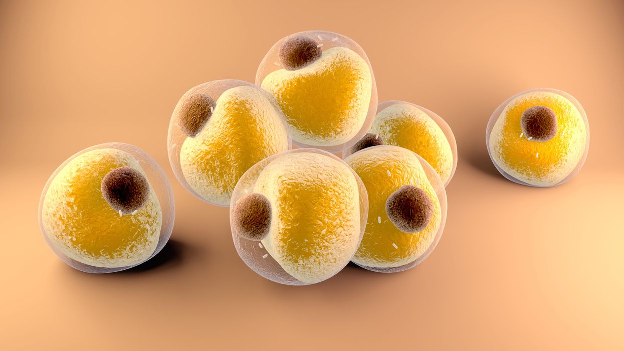
Now researchers have found a more direct reason: SARS-CoV-2, the virus that causes Covid-19, can infect adipocytes, more commonly known as fat cells, and macrophages, immune cells that are part of the broader matrix of cells that support fat tissue. Stanford University researchers Catherine Blish and Tracey McLaughlin are senior authors of the study.
Most of us think of fat as the spare tire that can accumulate around the middle as we age, but fat also is present closer to most internal organs. McLaughlin's research has focused on epicardial fat, “which sits right on top of the heart with no physical barrier at all,” she says. So if that fat got infected and inflamed, it might directly affect the heart.” That could help explain cardiovascular problems associated with Covid-19 infections.
Looking at tissue taken from autopsy, there was evidence of SARS-CoV-2 virus inside the fat cells as well as surrounding inflammation. In fat cells and immune cells harvested from health humans, infection in the laboratory drove "an inflammatory response, particularly in the macrophages…They secreted proteins that are typically seen in a cytokine storm” where the immune response runs amok with potential life-threatening consequences. This suggests to McLaughlin “that there could be a regional and even a systemic inflammatory response following infection in fat.”
It is easy to see how the airborne SARS-CoV-2 virus infects the nose and lungs, but how does it get into fat tissue? That is a mystery and the source of ample speculation.
The macrophages studied by McLaughlin and Blish were spewing out inflammatory proteins, While the the virus within them was replicating, the new viral particles were not able to replicate within those cells. It was a different story in the fat cells. “When [the virus] gets into the fat cells, it not only replicates, it's a productive infection, which means the resulting viral particles can infect another cell,” including microphages, McLaughlin explains. It seems to be a symbiotic tango of the virus between the two cell types that keeps the cycle going.
It is easy to see how the airborne SARS-CoV-2 virus infects the nose and lungs, but how does it get into fat tissue? That is a mystery and the source of ample speculation.
Macrophages are mobile; they engulf and carry invading pathogens to lymphoid tissue in the lymph nodes, tonsils and elsewhere in the body to alert T cells of the immune system to the pathogen. Perhaps some of them also carry the virus through the bloodstream to more distant tissue.
ACE2 receptors are the means by which SARS-CoV-2 latches on to and enters most cells. They are not thought to be common on fat cells, so initially most researchers thought it unlikely they would become infected.
However, while some cell receptors always sit on the surface of the cell, other receptors are expressed on the surface only under certain conditions. Philipp Scherer, a professor of internal medicine and director of the Touchstone Diabetes Center at the University of Texas Southwestern Medical Center, suggests that, in people who have obesity, “There might be higher levels of dysfunctional [fat cells] that facilitate entry of the virus,” either through transiently expressed ACE2 or other receptors. Inflammatory proteins generated by macrophages might contribute to this process.
Another hypothesis is that viral RNA might be smuggled into fat cells as cargo in small bits of material called extracellular vesicles, or EVs, that can travel between cells. Other researchers have shown that when EVs express ACE2 receptors, they can act as decoys for SARS-CoV-2, where the virus binds to them rather than a cell. These scientists are working to create drugs that mimic this decoy effect as an approach to therapy.
Do fat cells play a role in Long Covid? “Fat cells are a great place to hide. You have all the energy you need and fat cells turn over very slowly; they have a half-life of ten years,” says Scherer. Observational studies suggest that acute Covid-19 can trigger the onset of diabetes especially in people who are overweight, and that patients taking medicines to regulate their diabetes “were actually quite protective” against acute Covid-19. Scherer has funding to study the risks and benefits of those drugs in animal models of Long Covid.
McLaughlin says there are two areas of potential concern with fat tissue and Long Covid. One is that this tissue might serve as a “big reservoir where the virus continues to replicate and is sent out” to other parts of the body. The second is that inflammation due to infected fat cells and macrophages can result in fibrosis or scar tissue forming around organs, inhibiting their function. Once scar tissue forms, the tissue damage becomes more difficult to repair.
Current Covid-19 treatments work by stopping the virus from entering cells through the ACE2 receptor, so they likely would have no effect on virus that uses a different mechanism. That means another approach will have to be developed to complement the treatments we already have. So the best advice McLaughlin can offer today is to keep current on vaccinations and boosters and lose weight to reduce the risk associated with obesity.

