CRISPR base editing gives measure of hope to people with muscular dystrophy

Next year, a neurologist will test CRISPR base editing in a trial of five people with muscular dystrophy to see if their muscles accept corrected cells and whether they multiply and take over the function of damaged cells.
When Martin Weber climbs the steps to his apartment on the fifth floor in Munich, an attentive observer might notice that he walks a little unevenly. “That’s because my calf muscles were the first to lose strength,” Weber explains.
About three years ago, the now 19-year-old university student realized that he suddenly had trouble keeping up with his track team at school. At tennis tournaments, he seemed to lose stamina after the first hour. “But it was still within the norm,” he says. “So it took a while before I noticed something was seriously wrong.” A blood test showed highly elevated liver markers. His parents feared he had liver cancer until a week-long hospital visit and scores of tests led to a diagnosis: hereditary limb-girdle muscular dystrophy, an incurable genetic illness that causes muscles to deteriorate.
As you read this text, you will surely use several muscles without being aware of them: Your heart muscle pumps blood through your arteries, your eye muscles let you follow the words in this sentence, and your hand muscles hold the tablet or cell phone. Muscles make up 40 percent of your body weight; we usually have 656 of them. Now imagine they are slowly losing their strength. No training, no protein shake can rebuild their function.
This is the reality for most people in Simone Spuler’s outpatient clinic at the Charité Hospital in Berlin, Germany: Almost all of her 2,500 patients have muscular dystrophy, a progressive illness striking mostly young people. Muscle decline leads to a wheelchair and, eventually, an early death due to a heart attack or the inability to breathe. In Germany alone, 300,000 people live with this illness, the youngest barely a year old. The CDC estimates that its most common form, Duchenne, affects 1 in every 3,500 to 6,000 male births each year in the United States.
The devastating progression of the disease is what motivates Spuler and her team of 25 scientists to find a cure. In 2019, they made a spectacular breakthrough: For the first time, they successfully used mRNA to introduce the CRISPR-Cas9 tool into human muscle stem cells to repair the dystrophy. “It’s really just one tiny molecule that doesn’t work properly,” Spuler explains.
CRISPR-Cas9 is a technology that lets scientists select and alter parts of the genome. It’s still comparatively new but has advanced quickly since its discovery in the early 2010s. “We now have the possibility to repair certain mutations with genetic editing,” Spuler says. “It’s pure magic.”
She projects a warm, motherly air and a professional calm that inspires trust from her patients. She needs these qualities because the 60-year-old neurologist has one of the toughest jobs in the world: All day long, patients with the incurable diagnosis of muscular dystrophy come to her clinic, and she watches them decline over the years. “Apart from physiotherapy, there is nothing we can recommend right now,” she says. That motivated her early in her career, when she met her first patients at the Max Planck Institute for Neurobiology near Munich in the 1990s. “I knew I had 30, 40 years to find something.”
She learned from the luminaries of her profession with postdocs at the University of California San Diego, Harvard and Johns Hopkins, before serving as a clinical fellow at the Mayo Clinic. In 2005, the Charité offered her the opportunity to establish a specialized clinic for myasthenia, or muscular weakness. An important influence on Spuler, she says, has been the French microbiologist Emmanuelle Charpentier, who received the Nobel Prize in 2020 along with Jennifer Doudna for their CRISPR research, and has worked in Berlin since 2015.
When CRISPR was first introduced, it was mainly used to cut through DNA. However, the cut can lead to undesired side effects. For the muscle stem cells, Spuler now uses a base editor to repair the damaged molecule with super fine scissors or tweezers.

“Apart from physiotherapy, there is nothing we can recommend right now,” Spuler says about her patients with limb-girdle muscular dystrophy.
Pablo Castagnola
Last year, she proved that the method works in mice. Injecting repaired cells into the rodents led to new muscle fibers and, in 2021 and 2022, she passed the first safety meetings with the Paul-Ehrlich Institute, which is responsible for approving human gene editing trials in Germany. She raised the nearly four million Euros needed to test the new method in the first clinical trial in humans with limb-girdle muscular dystrophy, beginning with one muscle that can easily be measured, such as the biceps.
This spring, Weber and his parents drove the 400 miles from Munich to Berlin. At Spuler’s lab, her team took a biopsy from muscles in his left arm. The first two steps – extraction and repair in a culture dish – went according to plan; Spuler was able to repair the mutation in Weber’s cells outside his body.
Next year, Weber will be the youngest participant when Spuler starts to test the method in a trial of five people “in vivo,” inside their bodies. This will be the real moment of truth: Will the participants’ muscles accept the corrected cells? Will the cells multiply and take over the function of damaged cells, just like Spuler was able to do in her lab with the rodents?
The effort is costly and complex. “The biggest challenge is to make absolutely sure that we don’t harm the patient,” Spuler says. This means scanning their entire genomes, “so we don’t accidentally damage or knock out an important gene.”
Weber, who asked not to be identified by his real name, is looking forward to the trial and he feels confident that “the risks are comparatively small because the method will only be applied to one muscle. The worst that can happen is that it doesn’t work. But in the best case, the muscle function will improve.”
He was so impressed with the Charité scientists that he decided to study biology at his university. He’s read extensively about CRISPR, so he understands why he has three healthy siblings. “That’s the statistics,” the biologist in training explains. “You get two sets of genes from each parent, and you have to get two faulty mutations to have muscular dystrophy. So we fit the statistics exactly: One of us four kids inherited the mutation.”
It was his mother, a college teacher, and father, a physicist by training, who heard about Spuler’s research. Even though Weber does not live at home anymore, having a chronically ill son is nearly a full-time job for his mother, Annette. The Berlin visit and the trial are financed separately through private sponsors, but the fights with Weber’s health insurance are frustrating and time-consuming. “Physiotherapy is the only thing that helps a bit,” Weber says, “and yet, they fought us on approving it every step of the way.”
Spuler does not want to evoke unrealistic expectations. “Patients who are wheelchair-bound won’t suddenly get up and walk."
Her son continues to exercise as much as possible. Riding his bicycle to the university has become too difficult, so he got an e-scooter. He had to give up competitive tennis because he does not have the stamina for a two-hour match, but he can still play with his dad or his buddies for an hour. His closest friends know about the diagnosis. “They help me, for instance, to lift something heavy because I can’t do that anymore,” Weber says.
The family was elated to find medical support at the Munich Muscle Center by the German Alliance for Muscular Patients and then at Spuler’s clinic in Berlin. “When you hear that this is a progressive illness with no chance of improvement, your world collapses as a parent,” Annette Weber says. “And then all of a sudden, there is this woman who sees scientific progress as an opportunity. Even just to be able to participate in the study is fantastic.”
Spuler does not want to evoke unrealistic expectations. “Patients who are wheelchair-bound won’t suddenly get up and walk,” she says. After all, she will start by applying the gene editor to only one muscle, “but it would be a big step if even a small muscle that is essential to grip something, or to swallow, regains function.”
Weber agrees. “I understand that I won’t regain 100 percent of my muscle function but even a small improvement or at least halting the deterioration is the goal.”
And yet, Spuler and others are ultimately searching for a true solution. In a separate effort, Massachusetts-based biotech company Sarepta announced this month it will seek expedited regulators’ approval to treat Duchenne patients with its investigational gene therapy. Unlike Spuler’s methods, Sarepta focuses specifically on the Duchenne form of muscular dystrophy, and it uses an adeno-assisted virus to deliver the therapy.
Spuler’s vision is to eventually apply gene editing to the entire body of her patients. To speed up the research, she and a colleague founded a private research company, Myopax. If she is able to prove that the body accepts the edited cells, the technique could be used for other monogenetic illnesses as well. “When we speak of genetic editing, many are scared and say, oh no, this is God’s work,” says Spuler. But she sees herself as a mechanic, not a divine being. “We really just exchange a molecule, that’s it.”
If everything goes well, Weber hopes that ten years from now, he will be the one taking biopsies from the next generation of patients and repairing their genes.
Indigenous wisdom plus honeypot ants could provide new antibiotics
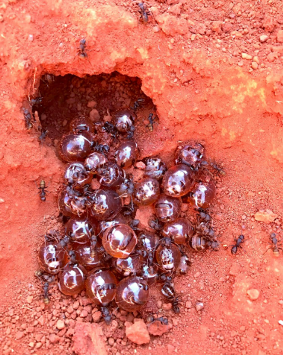
Indigenous people in Australia dig pits next to a honeypot colony. Scientists think the honey can be used to make new antimicrobial drugs.
For generations, the Indigenous Tjupan people of Australia enjoyed the sweet treat of honey made by honeypot ants. As a favorite pastime, entire families would go searching for the underground colonies, first spotting a worker ant and then tracing it to its home. The ants, which belong to the species called Camponotus inflatus, usually build their subterranean homes near the mulga trees, Acacia aneura. Having traced an ant to its tree, it would be the women who carefully dug a pit next to a colony, cautious not to destroy the entire structure. Once the ant chambers were exposed, the women would harvest a small amount to avoid devastating the colony’s stocks—and the family would share the treat.
The Tjupan people also knew that the honey had antimicrobial properties. “You could use it for a sore throat,” says Danny Ulrich, a member of the Tjupan nation. “You could also use it topically, on cuts and things like that.”
These hunts have become rarer, as many of the Tjupan people have moved away and, up until now, the exact antimicrobial properties of the ant honey remained unknown. But recently, scientists Andrew Dong and Kenya Fernandes from the University of Sydney, joined Ulrich, who runs the Honeypot Ants tours in Kalgoorlie, a city in Western Australia, on a honey-gathering expedition. Afterwards, they ran a series of experiments analyzing the honey’s antimicrobial activity—and confirmed that the Indigenous wisdom was true. The honey was effective against Staphylococcus aureus, a common pathogen responsible for sore throats, skin infections like boils and sores, and also sepsis, which can result in death. Moreover, the honey also worked against two species of fungi, Cryptococcus and Aspergillus, which can be pathogenic to humans, especially those with suppressed immune systems.
In the era of growing antibiotic resistance and the rising threat of pathogenic fungi, these findings may help scientists identify and make new antimicrobial compounds. “Natural products have been honed over thousands and millions of years by nature and evolution,” says Fernandes. “And some of them have complex and intricate properties that make them really important as potential new antibiotics. “

In an era of growing resistance to antibiotics and new threats of fungi infections, the latest findings about honeypot ants are helping scientists identify new antimicrobial drugs.
Danny Ulrich
Bee honey is also known for its antimicrobial properties, but bees produce it very differently than the ants. Bees collect nectar from flowers, which they regurgitate at the hive and pack into the hexagonal honeycombs they build for storage. As they do so, they also add into the mix an enzyme called glucose oxidase produced by their glands. The enzyme converts atmospheric oxygen into hydrogen peroxide, a reactive molecule that destroys bacteria and acts as a natural preservative. After the bees pack the honey into the honeycombs, they fan it with their wings to evaporate the water. Once a honeycomb is full, the bees put a beeswax cover on it, where it stays well-preserved thanks to the enzymatic action, until the bees need it.
Less is known about the chemistry of ants’ honey-making. Similarly to bees, they collect nectar. They also collect the sweet sap of the mulga tree. Additionally, they also “milk” the aphids—small sap-sucking insects that live on the tree. When ants tickle the aphids with their antennae, the latter release a sweet substance, which the former also transfer to their colonies. That’s where the honey management difference becomes really pronounced. The ants don’t build any kind of structures to store their honey. Instead, they store it in themselves.
The workers feed their harvest to their fellow ants called repletes, stuffing them up to the point that their swollen bellies outgrow the ants themselves, looking like amber-colored honeypots—hence the name. Because of their size, repletes don’t move, but hang down from the chamber’s ceiling, acting as living feedstocks. When food becomes scarce, they regurgitate their reserves to their colony’s brethren. It’s not clear whether the repletes die afterwards or can be restuffed again. “That's a good question,” Dong says. “After they've been stretched, they can't really return to exactly the same shape.”
These replete ants are the “treat” the Tjupan women dug for. Once they saw the round-belly ants inside the chambers, they would reach in carefully and get a few scoops of them. “You see a lot of honeypot ants just hanging on the roof of the little openings,” says Ulrich’s mother, Edie Ulrich. The women would share the ants with family members who would eat them one by one. “They're very delicate,” shares Edie Ulrich—you have to take them out carefully, so they don’t accidentally pop and become a wasted resource. “Because you’d lose all this precious honey.”
Dong stumbled upon the honeypot ants phenomenon because he was interested in Indigenous foods and went on Ulrich’s tour. He quickly became fascinated with the insects and their role in the Indigenous culture. “The honeypot ants are culturally revered by the Indigenous people,” he says. Eventually he decided to test out the honey’s medicinal qualities.
The researchers were surprised to see that even the smallest, eight percent concentration of honey was able to arrest the growth of S. aureus.
To do this, the two scientists first diluted the ant honey with water. “We used something called doubling dilutions, which means that we made 32 percent dilutions, and then we halve that to 16 percent and then we half that to eight percent,” explains Fernandes. The goal was to obtain as much results as possible with the meager honey they had. “We had very, very little of the honeypot ant honey so we wanted to maximize the spectrum of results we can get without wasting too much of the sample.”
After that, the researchers grew different microbes inside a nutrient rich broth. They added the broth to the different honey dilutions and incubated the mixes for a day or two at the temperature favorable to the germs’ growth. If the resulting solution turned turbid, it was a sign that the bugs proliferated. If it stayed clear, it meant that the honey destroyed them. The researchers were surprised to see that even the smallest, eight percent concentration of honey was able to arrest the growth of S. aureus. “It was really quite amazing,” Fernandes says. “Eight milliliters of honey in 92 milliliters of water is a really tiny amount of honey compared to the amount of water.”
Similar to bee honey, the ants’ honey exhibited some peroxide antimicrobial activity, researchers found, but given how little peroxide was in the solution, they think the honey also kills germs by a different mechanism. “When we measured, we found that [the solution] did have some hydrogen peroxide, but it didn't have as much of it as we would expect based on how active it was,” Fernandes says. “Whether this hydrogen peroxide also comes from glucose oxidase or whether it's produced by another source, we don't really know,” she adds. The research team does have some hypotheses about the identity of this other germ-killing agent. “We think it is most likely some kind of antimicrobial peptide that is actually coming from the ant itself.”
The honey also has a very strong activity against the two types of fungi, Cryptococcus and Aspergillus. Both fungi are associated with trees and decaying leaves, as well as in the soils where ants live, so the insects likely have evolved some natural defense compounds, which end up inside the honey.
It wouldn’t be the first time when modern medicines take their origin from the natural world or from the indigenous people’s knowledge. The bark of the cinchona tree native to South America contains quinine, a substance that treats malaria. The Indigenous people of the Andes used the bark to quell fever and chills for generations, and when Europeans began to fall ill with malaria in the Amazon rainforest, they learned to use that medicine from the Andean people.
The wonder drug aspirin similarly takes its origin from a bark of a tree—in this case a willow.
Even some anticancer compounds originated from nature. A chemotherapy drug called Paclitaxel, was originally extracted from the Pacific yew trees, Taxus brevifolia. The samples of the Pacific yew bark were first collected in 1962 by researchers from the United States Department of Agriculture who were looking for natural compounds that might have anti-tumor activity. In December 1992, the FDA approved Paclitaxel (brand name Taxol) for the treatment of ovarian cancer and two years later for breast cancer.
In the era when the world is struggling to find new medicines fast enough to subvert a fungal or bacterial pandemic, these discoveries can pave the way to new therapeutics. “I think it's really important to listen to indigenous cultures and to take their knowledge because they have been using these sources for a really, really long time,” Fernandes says. Now we know it works, so science can elucidate the molecular mechanisms behind it, she adds. “And maybe it can even provide a lead for us to develop some kind of new treatments in the future.”
Lina Zeldovich has written about science, medicine and technology for Popular Science, Smithsonian, National Geographic, Scientific American, Reader’s Digest, the New York Times and other major national and international publications. A Columbia J-School alumna, she has won several awards for her stories, including the ASJA Crisis Coverage Award for Covid reporting, and has been a contributing editor at Nautilus Magazine. In 2021, Zeldovich released her first book, The Other Dark Matter, published by the University of Chicago Press, about the science and business of turning waste into wealth and health. You can find her on http://linazeldovich.com/ and @linazeldovich.
Blood Test Can Detect Lymphoma Cells Before a Tumor Grows Back
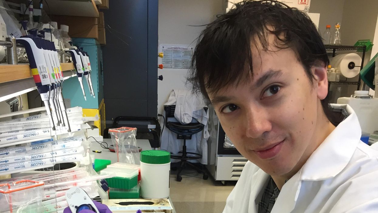
David Kurtz making DNA sequencing libraries in his lab.
When David M. Kurtz was doing his clinical fellowship at Stanford University Medical Center in 2009, specializing in lymphoma treatments, he found himself grappling with a question no one could answer. A typical regimen for these blood cancers prescribed six cycles of chemotherapy, but no one knew why. "The number seemed to be drawn out of a hat," Kurtz says. Some patients felt much better after just two doses, but had to endure the toxic effects of the entire course. For some elderly patients, the side effects of chemo are so harsh, they alone can kill. Others appeared to be cancer-free on the CT scans after the requisite six but then succumbed to it months later.
"Anecdotally, one patient decided to stop therapy after one dose because he felt it was so toxic that he opted for hospice instead," says Kurtz, now an oncologist at the center. "Five years down the road, he was alive and well. For him, just one dose was enough." Others would return for their one-year check up and find that their tumors grew back. Kurtz felt that while CT scans and MRIs were powerful tools, they weren't perfect ones. They couldn't tell him if there were any cancer cells left, stealthily waiting to germinate again. The scans only showed the tumor once it was back.
Blood cancers claim about 68,000 people a year, with a new diagnosis made about every three minutes, according to the Leukemia Research Foundation. For patients with B-cell lymphoma, which Kurtz focuses on, the survival chances are better than for some others. About 60 percent are cured, but the remaining 40 percent will relapse—possibly because they will have a negative CT scan, but still harbor malignant cells. "You can't see this on imaging," says Michael Green, who also treats blood cancers at University of Texas MD Anderson Medical Center.
The new blood test is sensitive enough to spot one cancerous perpetrator amongst one million other DNA molecules.
Kurtz wanted a better diagnostic tool, so he started working on a blood test that could capture the circulating tumor DNA or ctDNA. For that, he needed to identify the specific mutations typical for B-cell lymphomas. Working together with another fellow PhD student Jake Chabon, Kurtz finally zeroed-in on the tumor's genetic "appearance" in 2017—a pair of specific mutations sitting in close proximity to each other—a rare and telling sign. The human genome contains about 3 billion base pairs of nucleotides—molecules that compose genes—and in case of the B-cell lymphoma cells these two mutations were only a few base pairs apart. "That was the moment when the light bulb went on," Kurtz says.
The duo formed a company named Foresight Diagnostics, focusing on taking the blood test to the clinic. But knowing the tumor's mutational signature was only half the process. The other was fishing the tumor's DNA out of patients' bloodstream that contains millions of other DNA molecules, explains Chabon, now Foresight's CEO. It would be like looking for an escaped criminal in a large crowd. Kurtz and Chabon solved the problem by taking the tumor's "mug shot" first. Doctors would take the biopsy pre-treatment and sequence the tumor, as if taking the criminal's photo. After treatments, they would match the "mug shot" to all DNA molecules derived from the patient's blood sample to see if any molecular criminals managed to escape the chemo.
Foresight isn't the only company working on blood-based tumor detection tests, which are dubbed liquid biopsies—other companies such as Natera or ArcherDx developed their own. But in a recent study, the Foresight team showed that their method is significantly more sensitive in "fishing out" the cancer molecules than existing tests. Chabon says that this test can detect circulating tumor DNA in concentrations that are nearly 100 times lower than other methods. Put another way, it's sensitive enough to spot one cancerous perpetrator amongst one million other DNA molecules.
They also aim to extend their test to detect other malignancies such as lung, breast or colorectal cancers.
"It increases the sensitivity of detection and really catches most patients who are going to progress," says Green, the University of Texas oncologist who wasn't involved in the study, but is familiar with the method. It would also allow monitoring patients during treatment and making better-informed decisions about which therapy regimens would be most effective. "It's a minimally invasive test," Green says, and "it gives you a very high confidence about what's going on."
Having shown that the test works well, Kurtz and Chabon are planning a new trial in which oncologists would rely on their method to decide when to stop or continue chemo. They also aim to extend their test to detect other malignancies such as lung, breast or colorectal cancers. The latest genome sequencing technologies have sequenced and catalogued over 2,500 different tumor specimens and the Foresight team is analyzing this data, says Chabon, which gives the team the opportunity to create more molecular "mug shots."
The team hopes that that their blood cancer test will become available to patients within about five years, making doctors' job easier, and not only at the biological level. "When I tell patients, "good news, your cancer is in remission', they ask me, 'does it mean I'm cured?'" Kurtz says. "Right now I can't answer this question because I don't know—but I would like to." His company's test, he hopes, will enable him to reply with certainty. He'd very much like to have the power of that foresight.
This article is republished from our archives to coincide with Blood Cancer Awareness Month, which highlights progress in cancer diagnostics and treatment.
Lina Zeldovich has written about science, medicine and technology for Popular Science, Smithsonian, National Geographic, Scientific American, Reader’s Digest, the New York Times and other major national and international publications. A Columbia J-School alumna, she has won several awards for her stories, including the ASJA Crisis Coverage Award for Covid reporting, and has been a contributing editor at Nautilus Magazine. In 2021, Zeldovich released her first book, The Other Dark Matter, published by the University of Chicago Press, about the science and business of turning waste into wealth and health. You can find her on http://linazeldovich.com/ and @linazeldovich.

