
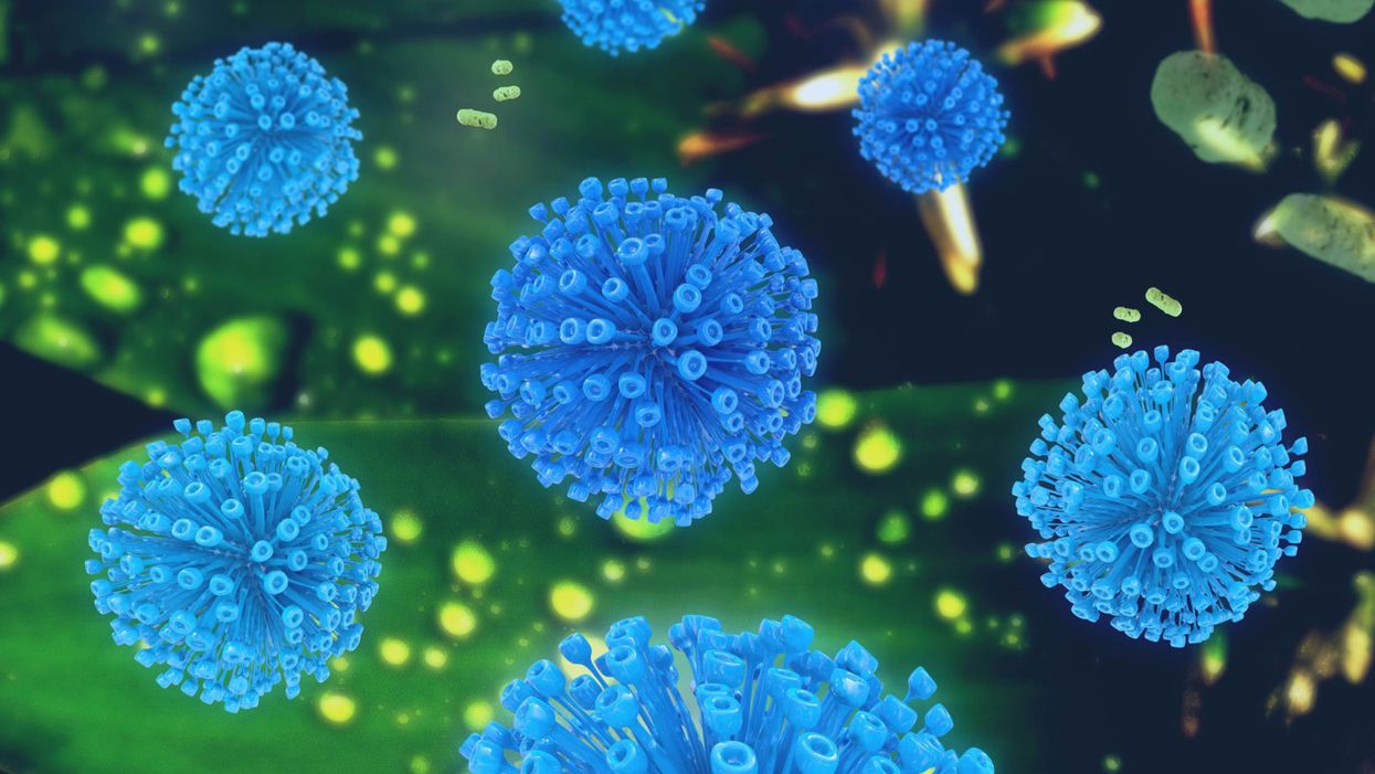
A retrovirus illustration.
Even with groundbreaking advances in cancer treatment and research over the past two centuries, the problem remains that some cancer does not respond to treatment. A subset of patients experience recurrence or metastasis, even when the original tumor is detected at an early stage.
"Why do some tumors evolve into metastatic disease that is then capable of spreading, while other tumors do not?"
Moreover, doctors are not able to tell in advance which patients will respond to treatment and which will not. This means that many patients endure conventional cancer therapies, like countless rounds of chemo and radiation, that do not ultimately increase their likelihood of survival.
Researchers are beginning to understand why some tumors respond to treatment and others do not. The answer appears to lie in the strange connection between human life at its earliest stages — and retroviruses. A retrovirus is different than a regular virus in that its RNA is reverse-transcribed into DNA, which makes it possible for its genetic material to be integrated into a host's genome, and passed on to subsequent generations.
Researchers have shown that reactivation of retroviral sequences is associated with the survival of developing embryos. Certain retroviral sequences must be expressed around the 8-cell stage for successful embryonic development. Active expression of retroviral sequences is required for proper functioning of human embryonic stem cells. These sequences must then shut down at the later state, or the embryo will fail to develop. And here's where things get really interesting: If specific stem cell-associated retroviral sequences become activated again later in life, they seem to play a role in some cancers becoming lethal.
"Eight to 10 million years ago, at the time when we became primates, the population was infected with a virus."
While some retroviral sequences in our genome contribute to the restriction of viral infection and appear to have contributed to the development of the placenta, they can also, if expressed at the wrong time, drive the development of cancer stem cells. Described as the "beating hearts" of treatment-resistant tumors, cancer stem cells are robust and long-living, and they can maintain the ability to proliferate indefinitely.
This apparent connection has inspired Gennadi V. Glinsky, a research scientist at the Institute of Engineering in Medicine at UC San Diego, to find better ways to diagnose and treat metastatic cancer. Glinsky specializes in the development of new technologies, methods, and system integration approaches for personalized genomics-guided prevention and precision therapy of cancer and other common human disorders. We spoke with him about his work and the exciting possibilities it may open up for cancer patients. This interview has been edited and condensed for clarity.
What key questions have driven your research in this area?
I was thinking for years that the major mysteries are: Why do some tumors evolve into metastatic disease that is then capable of spreading, while other tumors do not? What explains some cancer cells' ability to get into the blood or lymph nodes and be able to survive in this very foreign, hostile environment of circulatory channels, and then be able to escape and take root elsewhere in the body?
"If you detect conventional cancer early, and treat it early, it will be cured. But with cancer involving stem cells, even if you diagnose it early, it will come back."
When we were able to do genomic analysis on enough early stage cancers, we arrived at an alternative concept of cancer that starts in the stem cells. Stem cells exist throughout our bodies, so in the case of cancer starting in stem cells you will have metastatic properties … because that's what stem cells do. They can travel throughout the body, they can make any other type of cell or resemble them.
So there are basically two types of cancer: conventional non-stem cell cancer and stem cell-like cancer. If you detect conventional cancer early, and treat it early, it will be cured. But with cancer involving stem cells, even if you diagnose it early, it will come back.
What causes some cancer to originate in stem cells?
Cancer stem cells possess stemness [or the ability to self-renew, differentiate, and survive chemical and physical insults]. Stemness is driven by the reactivation of retroviral sequences that have been integrated into the human genome.
Tell me about these retroviral sequences.
Eight to 10 million years ago, at the time when we became primates, the population was infected with a virus. Part of the population survived and the virus was integrated into our primate ancestors' genome. These are known as human endogenous retroviruses, or HERVs. The DNA of the host cells became carriers of these retroviral sequences, and whenever the host cells multiply, they carry the sequences in them and pass them on to future generations.
This pattern of infection and integration of retroviral sequences has happened thousands of times during our evolutionary history. As a result, eight percent of the human genome is derived from these different retroviral sequences.
We've found that some HERVs are expressed in some cancers. For example, 10-15 percent of prostate cancer is stem cell-like. But at first it was not understood what this HERV expression meant.
Gennadi V. Glinsky, a research scientist at the Institute of Engineering in Medicine at UC San Diego.
(Courtesy)
How have you endeavored to solve this in your lab?
We were trying to track down metastatic prostate cancer. We found a molecular signature of prostate cancer that made the prostate tumors look like stem cells. And those were the ones likely to fail cancer therapy. Then we applied this signature to other types of cancers and we found that uniformly, tumors that exhibit stemness fail therapy.
Then in 2014, several breakthrough papers came out that linked the activation of the retroviral sequences in human embryonic stem cells and in human embryo development. When I read these papers, it occurred to me that if these retroviral sequences are required for pluripotency in human embryonic stem cells, they must be involved in stem cell-resembling human cancer that's likely to fail therapy.
What was one of the biggest aha moments in your cancer research?
Several major labs around the U.S. took advantage of The Cancer Genome Anatomy Project, which made it possible to have access to about 12,000 individual human tumors across a spectrum of 30 or so cancer types. This is the largest set of tumors that's ever been made available in a comprehensive and state of the art way. So we now know all there is to know about the genetics of these tumors, including the long-term clinical outcome.
"When we cross-referenced these 10,713 human cancer survival genes to see how many are part of the retroviral network in human cells, we found that the answer was 97 percent!"
These labs identified 10,713 human genes that were associated with the likelihood of patients surviving or dying after [cancer] treatment. I call them the human cancer survival genes, and there are two classes of them: one whose high expression in tumors correlates with an increased likelihood of survival and one whose high expression in tumors correlates with a decreased likelihood of survival.
When we cross-referenced these 10,713 human cancer survival genes to see how many are part of the retroviral network in human cells, we found that the answer was 97 percent!
How will all of this new knowledge change how cancer is treated?
To make cancer stem cells vulnerable to treatment, you need to interfere with stemness and the stemness network. And to do this, you would need to identify the retroviral component of the network, and interfere with this component therapeutically.
The real breakthrough will come when we start to treat these early stage stem cell-like cancers with stem cell-targeting therapy that we are trying to develop. And with our ability to detect the retroviral genome activation, we will be able to detect stem cell-like cancer very early on.
How far away are we from being able to apply this information clinically?
We have two molecule [treatment] candidates. We know that they efficiently interfere with the stemness program in the cells. The road to clinical trials is typically a long one, but since we're clear about our targets, it's a shorter road. We would like to say it's two to three years until we can start a human trial.
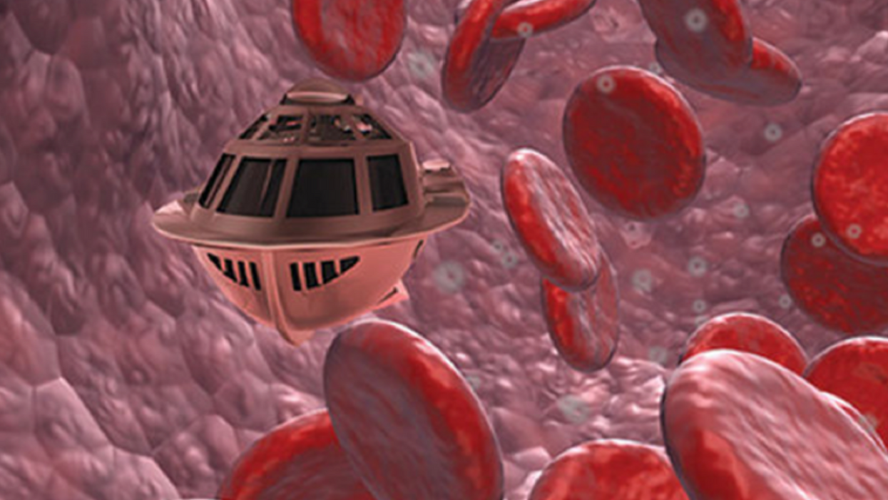
A movie still from the 1966 film "Fantastic Voyage"
"Chemotherapy is delivered systemically," Bionaut-founder and CEO Michael Shpigelmacher says. "Often only a small percentage arrives at the location where it is actually needed."
But what if it was possible to send a tiny robot through the body to attack a tumor or deliver a drug at exactly the right location?
Several startups and academic institutes worldwide are working to develop such a solution but Bionaut Labs seems the furthest along in advancing its invention. "You can think of the Bionaut as a tiny screw that moves through the veins as if steered by an invisible screwdriver until it arrives at the tumor," Shpigelmacher explains. Via Zoom, he shares the screen of an X-ray machine in his Culver City lab to demonstrate how the half-transparent, yellowish device winds its way along the spine in the body. The nanobot contains a tiny but powerful magnet. The "invisible screwdriver" is an external magnetic field that rotates that magnet inside the device and gets it to move and change directions.
The current model has a diameter of less than a millimeter. Shpigelmacher's engineers could build the miniature vehicle even smaller but the current size has the advantage of being big enough to see with bare eyes. It can also deliver more medicine than a tinier version. In the Zoom demonstration, the micorobot is injected into the spine, not unlike an epidural, and pulled along the spine through an outside magnet until the Bionaut reaches the brainstem. Depending which organ it needs to reach, it could be inserted elsewhere, for instance through a catheter.
"The hope is that we can develop a vehicle to transport medication deep into the body," says Max Planck scientist Tian Qiu.
Imagine moving a screw through a steak with a magnet — that's essentially how the device works. But of course, the Bionaut is considerably different from an ordinary screw: "At the right location, we give a magnetic signal, and it unloads its medicine package," Shpigelmacher says.
To start, Bionaut Labs wants to use its device to treat Parkinson's disease and brain stem gliomas, a type of cancer that largely affects children and teenagers. About 300 to 400 young people a year are diagnosed with this type of tumor. Radiation and brain surgery risk damaging sensitive brain tissue, and chemotherapy often doesn't work. Most children with these tumors live less than 18 months. A nanobot delivering targeted chemotherapy could be a gamechanger. "These patients really don't have any other hope," Shpigelmacher says.
Of course, the main challenge of the developing such a device is guaranteeing that it's safe. Because tissue is so sensitive, any mistake could risk disastrous results. In recent years, Bionaut has tested its technology in dozens of healthy sheep and pigs with no major adverse effects. Sheep make a good stand-in for humans because their brains and spines are similar to ours.
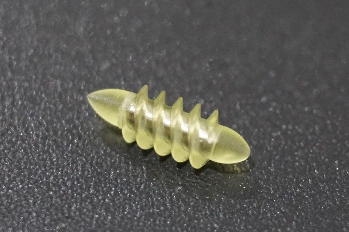
The Bionaut device is about the size of a grain of rice.
Bionaut Labs
"As the Bionaut moves through brain tissue, it creates a transient track that heals within a few weeks," Shpigelmacher says. The company is hoping to be the first to test a nanobot in humans. In December 2022, it announced that a recent round of funding drew $43.2 million, for a total of 63.2 million, enabling more research and, if all goes smoothly, human clinical trials by early next year.
Once the technique has been perfected, further applications could include addressing other kinds of brain disorders that are considered incurable now, such as Alzheimer's or Huntington's disease. "Microrobots could serve as a bridgehead, opening the gateway to the brain and facilitating precise access of deep brain structure – either to deliver medication, take cell samples or stimulate specific brain regions," Shpigelmacher says.
Robot-assisted hybrid surgery with artificial intelligence is already used in state-of-the-art surgery centers, and many medical experts believe that nanorobotics will be the instrument of the future. In 2016, three scientists were awarded the Nobel Prize in Chemistry for their development of "the world's smallest machines," nano "elevators" and minuscule motors. Since then, the scientific experiments have progressed to the point where applicable devices are moving closer to actually being implemented.
Bionaut's technology was initially developed by a research team lead by Peer Fischer, head of the independent Micro Nano and Molecular Systems Lab at the Max Planck Institute for Intelligent Systems in Stuttgart, Germany. Fischer is considered a pioneer in the research of nano systems, which he began at Harvard University more than a decade ago. He and his team are advising Bionaut Labs and have licensed their technology to the company.
"The hope is that we can develop a vehicle to transport medication deep into the body," says Max Planck scientist Tian Qiu, who leads the cooperation with Bionaut Labs. He agrees with Shpigelmacher that the Bionaut's size is perfect for transporting medication loads and is researching potential applications for even smaller nanorobots, especially in the eye, where the tissue is extremely sensitive. "Nanorobots can sneak through very fine tissue without causing damage."
In "Fantastic Voyage," Raquel Welch's adventures inside the body of a dissident scientist let her swim through his veins into his brain, but her shrunken miniature submarine is attacked by antibodies; she has to flee through the nerves into the scientist's eye where she escapes into freedom on a tear drop. In reality, the exit in the lab is much more mundane. The Bionaut simply leaves the body through the same port where it entered. But apart from the dramatization, the "Fantastic Voyage" was almost prophetic, or, as Shpigelmacher says, "Science fiction becomes science reality."
This article was first published by Leaps.org on April 12, 2021.
In 2013, the Human Brain Project set out to build a realistic computer model of the brain over ten years. Now, experts are reflecting on HBP's achievements with an eye toward the future.
Scholars have found that the project did help advance neuroscience more than some detractors initially expected, specifically in the area of brain simulations and virtual models. Using an interdisciplinary approach of combining technology, such as AI and digital simulations, with neuroscience, the HBP worked to gain a deeper understanding of the human brain’s complicated structure and functions, which in some cases led to novel treatments for brain disorders. Lastly, through online platforms, the HBP spearheaded a previously unmatched level of global neuroscience collaborations.
Simulating a human brain stirs up controversy
Right from the start, the project was plagued with controversy and condemnation. One of its prominent critics was Yves Fregnac, a professor in cognitive science at the Polytechnic Institute of Paris and research director at the French National Centre for Scientific Research. Fregnac argued in numerous articles that the HBP was overfunded based on proposals with unrealistic goals. “This new way of over-selling scientific targets, deeply aligned with what modern society expects from mega-sciences in the broad sense (big investment, big return), has been observed on several occasions in different scientific sub-fields,” he wrote in one of his articles, “before invading the field of brain sciences and neuromarketing.”
"A human brain model can simulate an experiment a million times for many different conditions, but the actual human experiment can be performed only once or a few times," said Viktor Jirsa, a professor at Aix-Marseille University.
Responding to such critiques, the HBP worked to restructure the effort in its early days with new leadership, organization, and goals that were more flexible and attainable. “The HBP got a more versatile, pluralistic approach,” said Viktor Jirsa, a professor at Aix-Marseille University and one of the HBP lead scientists. He believes that these changes fixed at least some of HBP’s issues. “The project has been on a very productive and scientifically fruitful course since then.”
After restructuring, the HBP became a European hub on brain research, with hundreds of scientists joining its growing network. The HBP created projects focused on various brain topics, from consciousness to neurodegenerative diseases. HBP scientists worked on complex subjects, such as mapping out the brain, combining neuroscience and robotics, and experimenting with neuromorphic computing, a computational technique inspired by the human brain structure and function—to name just a few.
Simulations advance knowledge and treatment options
In 2013, it seemed that bringing neuroscience into a digital age would be farfetched, but research within the HBP has made this achievable. The virtual maps and simulations various HBP teams create through brain imaging data make it easier for neuroscientists to understand brain developments and functions. The teams publish these models on the HBP’s EBRAINS online platform—one of the first to offer access to such data to neuroscientists worldwide via an open-source online site. “This digital infrastructure is backed by high-performance computers, with large datasets and various computational tools,” said Lucy Xiaolu Wang, an assistant professor in the Resource Economics Department at the University of Massachusetts Amherst, who studies the economics of the HBP. That means it can be used in place of many different types of human experimentation.
Jirsa’s team is one of many within the project that works on virtual brain models and brain simulations. Compiling patient data, Jirsa and his team can create digital simulations of different brain activities—and repeat these experiments many times, which isn’t often possible in surgeries on real brains. “A human brain model can simulate an experiment a million times for many different conditions,” Jirsa explained, “but the actual human experiment can be performed only once or a few times.” Using simulations also saves scientists and doctors time and money when looking at ways to diagnose and treat patients with brain disorders.
Compiling patient data, scientists can create digital simulations of different brain activities—and repeat these experiments many times.
The Human Brain Project
Simulations can help scientists get a full picture that otherwise is unattainable. “Another benefit is data completion,” added Jirsa, “in which incomplete data can be complemented by the model. In clinical settings, we can often measure only certain brain areas, but when linked to the brain model, we can enlarge the range of accessible brain regions and make better diagnostic predictions.”
With time, Jirsa’s team was able to move into patient-specific simulations. “We advanced from generic brain models to the ability to use a specific patient’s brain data, from measurements like MRI and others, to create individualized predictive models and simulations,” Jirsa explained. He and his team are working on this personalization technique to treat patients with epilepsy. According to the World Health Organization, about 50 million people worldwide suffer from epilepsy, a disorder that causes recurring seizures. While some epilepsy causes are known others remain an enigma, and many are hard to treat. For some patients whose epilepsy doesn’t respond to medications, removing part of the brain where seizures occur may be the only option. Understanding where in the patients’ brains seizures arise can give scientists a better idea of how to treat them and whether to use surgery versus medications.
“We apply such personalized models…to precisely identify where in a patient’s brain seizures emerge,” Jirsa explained. “This guides individual surgery decisions for patients for which surgery is the only treatment option.” He credits the HBP for the opportunity to develop this novel approach. “The personalization of our epilepsy models was only made possible by the Human Brain Project, in which all the necessary tools have been developed. Without the HBP, the technology would not be in clinical trials today.”
Personalized simulations can significantly advance treatments, predict the outcome of specific medical procedures and optimize them before actually treating patients. Jirsa is watching this happen firsthand in his ongoing research. “Our technology for creating personalized brain models is now used in a large clinical trial for epilepsy, funded by the French state, where we collaborate with clinicians in hospitals,” he explained. “We have also founded a spinoff company called VB Tech (Virtual Brain Technologies) to commercialize our personalized brain model technology and make it available to all patients.”
The Human Brain Project created a level of interconnectedness within the neuroscience research community that never existed before—a network not unlike the brain’s own.
Other experts believe it’s too soon to tell whether brain simulations could change epilepsy treatments. “The life cycle of developing treatments applicable to patients often runs over a decade,” Wang stated. “It is still too early to draw a clear link between HBP’s various project areas with patient care.” However, she admits that some studies built on the HBP-collected knowledge are already showing promise. “Researchers have used neuroscientific atlases and computational tools to develop activity-specific stimulation programs that enabled paraplegic patients to move again in a small-size clinical trial,” Wang said. Another intriguing study looked at simulations of Alzheimer’s in the brain to understand how it evolves over time.
Some challenges remain hard to overcome even with computer simulations. “The major challenge has always been the parameter explosion, which means that many different model parameters can lead to the same result,” Jirsa explained. An example of this parameter explosion could be two different types of neurodegenerative conditions, such as Parkinson’s and Huntington’s diseases. Both afflict the same area of the brain, the basal ganglia, which can affect movement, but are caused by two different underlying mechanisms. “We face the same situation in the living brain, in which a large range of diverse mechanisms can produce the same behavior,” Jirsa said. The simulations still have to overcome the same challenge.
Understanding where in the patients’ brains seizures arise can give scientists a better idea of how to treat them and whether to use surgery versus medications.
The Human Brain Project
A network not unlike the brain’s own
Though the HBP will be closing this year, its legacy continues in various studies, spin-off companies, and its online platform, EBRAINS. “The HBP is one of the earliest brain initiatives in the world, and the 10-year long-term goal has united many researchers to collaborate on brain sciences with advanced computational tools,” Wang said. “Beyond the many research articles and projects collaborated on during the HBP, the online neuroscience research infrastructure EBRAINS will be left as a legacy even after the project ends.”
Those who worked within the HBP see the end of this project as the next step in neuroscience research. “Neuroscience has come closer to very meaningful applications through the systematic link with new digital technologies and collaborative work,” Jirsa stated. “In that way, the project really had a pioneering role.” It also created a level of interconnectedness within the neuroscience research community that never existed before—a network not unlike the brain’s own. “Interconnectedness is an important advance and prerequisite for progress,” Jirsa said. “The neuroscience community has in the past been rather fragmented and this has dramatically changed in recent years thanks to the Human Brain Project.”
According to its website, by 2023 HBP’s network counted over 500 scientists from over 123 institutions and 16 different countries, creating one of the largest multi-national research groups in the world. Even though the project hasn’t produced the in-silico brain as Markram envisioned it, the HBP created a communal mind with immense potential. “It has challenged us to think beyond the boundaries of our own laboratories,” Jirsa said, “and enabled us to go much further together than we could have ever conceived going by ourselves.”